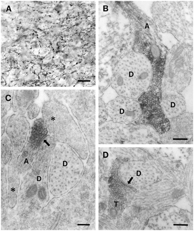
Light and electron microscopic characteristics of GAD65-immunoreactive fibers in P1 hippocampus. A, Light microscopy demonstrates numerous fine fibers with periodic swellings within the developing dendritic region of CA3. The remaining panels demonstrate ultrastructural features of such fibers in the various hippocampal fields. B, A GAD65-labeled axon-like process (A) contains a large number of vesicles and courses between several dendritic profiles (D) of the CA3 field. Immunoreactivity is highly concentrated in vesicle-filled regions of the process. C, GAD65 immunoreactivity is concentrated at a vesicle-filled site within an axon-like process (A) in the molecular layer of the dentate gyrus. This site is in close contact (arrow) with one of several dendritic profiles (D) in the region. The labeled process is also located in close proximity to several undifferentiated or developing neuronal profiles, which have a paucity of organelles (asterisks). D, Some immunolabeled profiles within the dendritic regions exhibit features of mature axon terminals (T), as illustrated by the high concentration of vesicles, the presence of a mitochondrial profile, and the establishment of a prototypic synaptic contact (arrow) with a dendrite (D) of the CA3 field. Scale bars: A, 10 μm; B, 0.3 μm; C, 0.2 μm; D, 0.3 μm.


