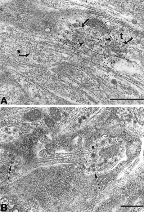
Fig. 2.
Glutamate-immunoreactive varicosities can be detected by electron microscopy in myenteric ganglia. A, Small, clear, round synaptic vesicles (arrow) are present in all glutamate-immunoreactive nerve terminals (t). The black dots(arrowhead) are gold particles (10 nm), which identify the axon as containing high levels of glutamate. A second terminal (*) also contains clear, round synaptic vesicles (arrow) but few gold particles. Note that there is some diffusion of the amino acid. B, Varicosities in which large, dense-cored vesicles (arrow) predominate are not labeled by antibodies to glutamate. Scale bar, 0.5 μm.


