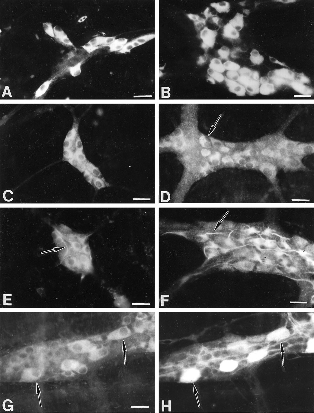
Differential distribution of AMPA receptor subunit immunoreactivity in the guinea pig small intestine. A, B, GluR1 immunoreactivity. A subset of neurons in the submucosal (A) and myenteric (B) plexus express GluR1 immunoreactivity. Immunoreactivity is concentrated in the cytoplasm. C, D, GluR2/3 immunoreactivity. The majority of neurons in the submucosal plexus (C) express GluR2/3 immunoreactivity. A subset of neurons in the myenteric plexus (D) expresses GluR2/3. These cells are small in size (arrow). E, F, GluR4 immunoreactivity. A subset of neurons in the submucosal (E) and myenteric (F) plexus express GluR4 immunoreactivity. Immunoreactivity is concentrated in the cytoplasm; however, proximal dendritic processes are also stained (arrow). G, H, GluR1 immunoreactivity (G) is expressed by the majority of calbindin-immunoreactive myenteric neurons (H, arrow). Scale bar, 30 μm.


