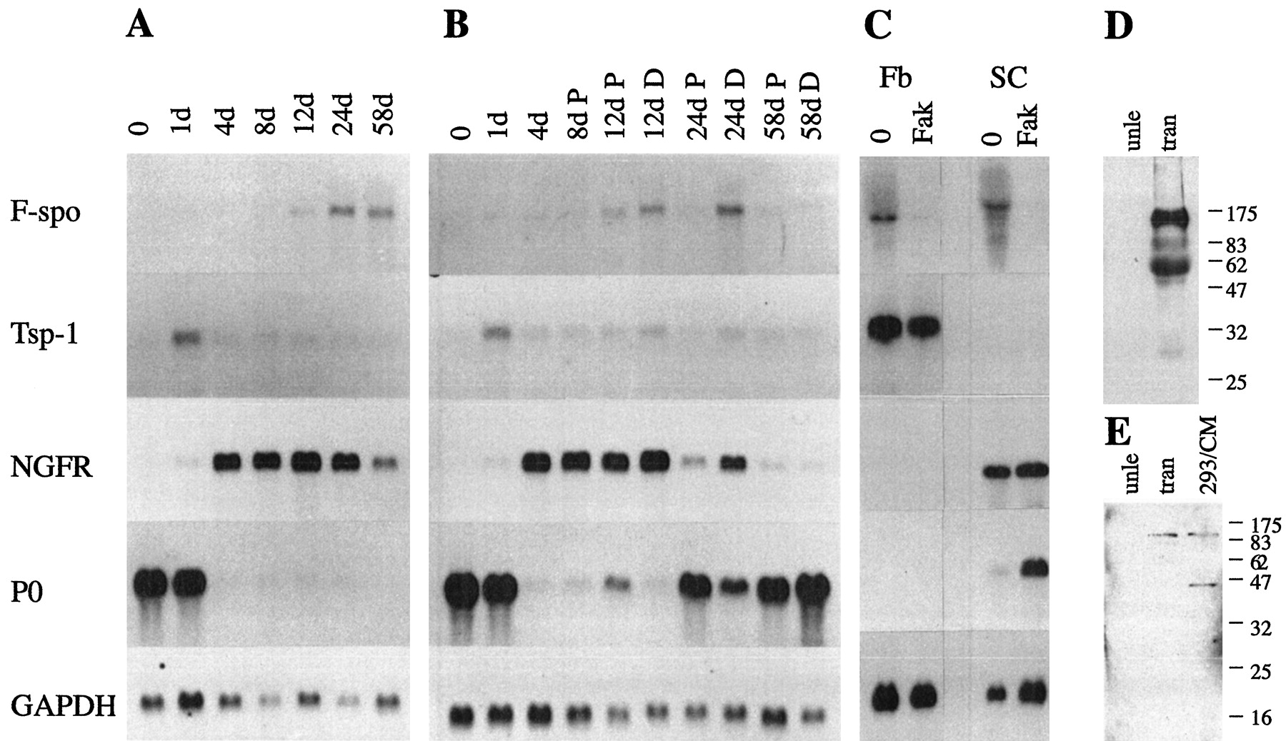
F-spondin mRNA and protein increases in lesioned adult rat sciatic nerve. Each lane in A–C contains an equal amount (10 μg) of total RNA isolated from the distal stumps of sciatic nerves that had been transected (A), crushed (B), or cultured Schwann cells and fibroblast (C). The number of days after each of these lesions is indicated; the 0 time point is from unlesioned nerves. The blots were successively hybridized with radiolabeled cDNA probes for F-spondin (F-spo), thrombospondin-1 (Tsp-1), NGFR,P0, and GAPDH mRNA and exposed to film for 2 weeks, 2 weeks, 1 d, 2 hr, and 3 d, respectively. B, The distal nerve stumps were divided into proximal (P) and distal (D) segments of equal lengths. C, RNA isolated from fibroblasts (Fb) or Schwann cells (SC) cultured for 3 d in DMEM + 10% FCS alone (0), or supplemented with 4 μm forskolin (Fak).D, E, Proteins were extracted from unlesioned adult rat sciatic nerves (unle) and the distal stumps of transected (tran) adult sciatic nerves 3 weeks after transection. Each lane contains an equal amount (4 μg) of protein extract. D, Western blot with the R5 Ab. A 115 and a 60 kDa protein are detected in the injured nerve. E, Western blot with the R2 Ab. A 115 kDa protein is present in the injured nerve, but the 40 kDa protein is not detected (compared with the F-spondin-transfected HEK 293 cells).


