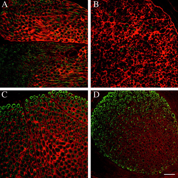
F-spondin immunoreactivity accumulates in the endoneurial ECM after nerve injury. Sections were double-labeled with R5 (TRITC) and myelin basic protein (FITC). Scale bar (shown inD): A–D, 10 μm. A, Transverse section of unlesioned adult rat sciatic nerve. F-spondin immunoreactivity is predominately localized in the ECM of fascicles composed of large myelinated fibers. B, Transverse section of an adult rat sciatic nerve 8 d after axotomy. F-spondin immunoreactivity is seen in the ECM, but at higher levels and in a more uniform distribution (no fascicle-to-fascicle variation) than in unlesioned nerve. C, Transverse section of unlesioned adult rat femoral motor branch. There is prominent F-spondin immunoreactivity in the ECM associated with the large, myelinated motor fibers. D, Transverse section of unlesioned adult rat femoral sensory branch (taken from the same section asC). The level of F-spondin immunoreactivity is less than that in the motor branch.


