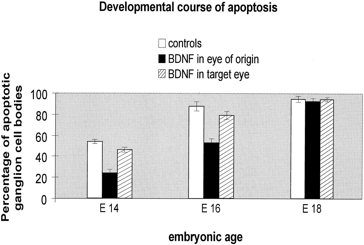
Fig. 8.
Graph corroborating the proportion of apoptotic cells at three typical embryonic days of investigation in normal and BDNF-treated embryos. At all stages, BDNF reduced the proportion of apoptotic silhouettes but was unable to rescue cells beyond the period of cell death (E18). There was a significant difference (p < 0.01, Student’s ttest) between embryos that received BDNF in the retina analyzed at both P14 and P16 and those that received BDNF in the retina contralateral to analysis.


