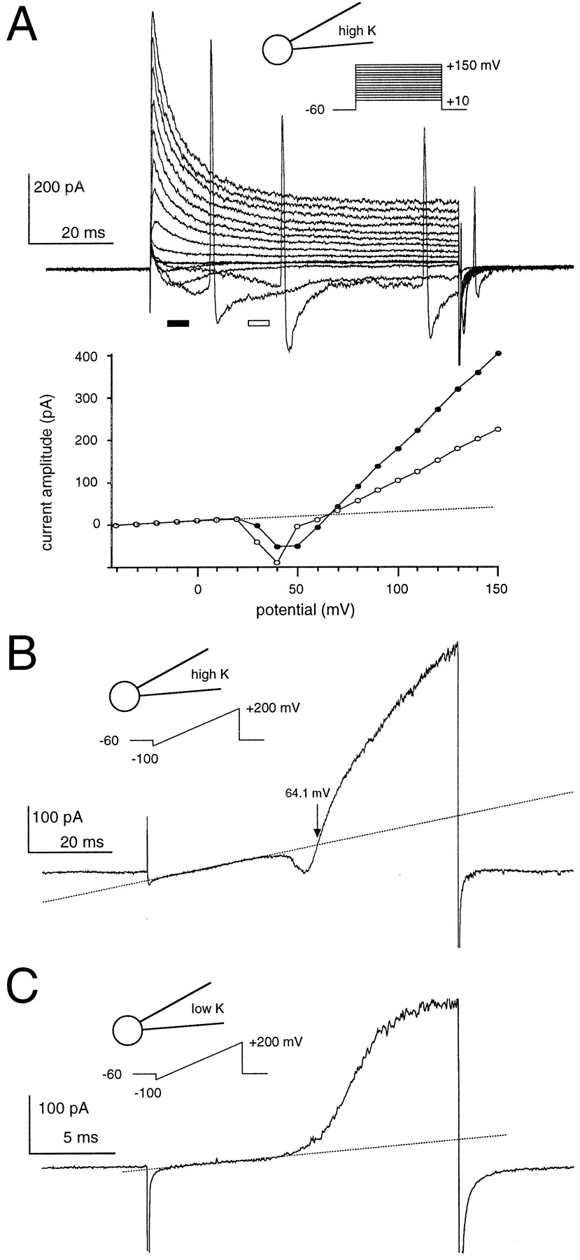
Voltage-gated K+ currents measured in cell-attached patches of CA1 interneurons. Ashows currents activated by applying depolarizing voltage steps, andB shows the response to a voltage ramp applied to the same cell, with a pipette solution containing 155 mmK+. A, The inward K+ current caused the generation of action currents in this cell (Lynch and Barry, 1989). At more depolarized potentials the current became outward and consisted of a transient and sustained component. Current–voltage curves (below), constructed from the average current amplitude at the times indicated by theblack and white bars in the top panel, show that the K+ current was superimposed on a linear leak (dotted lines) and reversed polarity from inward to outward between +60 and +70 mV. Note that the reversal potential of the voltage-gated current is independent of the relative contributions of the transient and sustained component.B, A voltage ramp stimulation gave a similarI–V profile, but no action currents were generated because of a reduced charge flux. With symmetrical K+, the current reversal at Vh = +64 mV indicated a membrane potential of −64 mV. C, With a low K+ pipette solution, in which 135 mmK+ was replaced with the impermeant cationN-methyl-d-glucamine, the voltage-gated current was outward for all voltages beyond the activation threshold, demonstrating its K+ selectivity.


