The Electrogenic Sodium Bicarbonate Cotransporter: Developmental Expression in Rat Brain and Possible Role in Acid Vulnerability
Abstract
The electrogenic sodium bicarbonate cotransporter (NBC) is expressed in glial cells in the brain and plays an important role in the regulation of both intracellular and extracellular pH. Differential vulnerability to acidosis between neurons and glia has been noted and may contribute to infarction after cerebral ischemia. Ionic substitution studies and inhibition of injury by 4,4′-di-isothiocyanostilbene-2,2′-disulfonic acid suggest that NBC is involved in astrocyte vulnerability to acidic injury. Recently two NBC cDNAs differing in 5′-untranslated and N-terminal coding sequence have been cloned from kidney and pancreas. We cloned one of these cDNAs from rat brain and demonstrate here that the clone is functional by expression in Xenopus oocytes. We determined the developmental and regional expression of NBC in the brain by in situ hybridization. Expression was observed in the spinal cord at embryonic day 17, whereas expression in brain was first seen at approximately postnatal day 0 (P0), increased at P15, and persisted in the adult brain. Expression was widespread throughout the cerebellum, cortex, olfactory bulb, and subcortical structures. Cellular resolution of the in situhybridization signal and double labeling for glial fibrillary acidic protein were consistent with a glial localization for NBC. Expression of NBC in 3T3 cells that do not normally express this transporter rendered them vulnerable to acid injury. The expression profile suggests that this transporter is critical during the later stages of brain development and could be one of the factors contributing to the different patterns of injury seen in perinatal versus adult cerebral ischemia.
Introduction
The electrogenic sodium bicarbonate cotransporter (NBC) has been studied extensively in glial cells from both invertebrates and vertebrates and from different brain regions (Deitmer and Schlue, 1989; Newman, 1991; O'Connor et al., 1994). The extracellular pH is an important determinant of neuronal excitability (Ransom, 1992). During physiological activation of neurons a shift to acidic pH occurs (Trapp et al., 1996), and modulation of the extracellular pH by astrocytes may be critical to setting the level of excitability of neurons (Ransom, 1992). During neuronal activity extracellular potassium increases, leading to glial depolarization and activation of the cotransporter (O'Connor et al., 1994). With activation of NBC the extracellular pH is acidified, while the intracellular pH of the astrocyte is alkalinized. The extracellular acidification may be instrumental in regulating local brain blood flow (Newman, 1991) in addition to its effect on neuronal excitability. This transporter plays an important role in physiological pH regulation and may play a critical role in pH regulation during pathophysiological events, such as brain ischemia (Lascola and Kraig, 1997).
Cerebral ischemia is associated with a fall in intracellular and extracellular pH (Kraig et al., 1986), and more severe acidosis correlates with more severe injury (Myers and Yamaguchi, 1977). However, in vitro, acidosis in the range seen in ischemia protects neurons from ischemia-like injury (Schurr et al., 1988;Giffard et al., 1990a; Tombaugh and Sapolsky, 1990) and attenuates glutamate-induced neuronal death (Giffard et al., 1990a). Surprisingly, astrocytes in vitro were more vulnerable to extracellular acidity-mediated injury than were neurons (Giffard et al., 1990b). Protection from hypoxic injury by acidity has been observed not only in neurons (Schurr et al., 1988; Giffard et al., 1990a) but also in cardiac (Bing et al., 1973), renal (Pentilla and Trump, 1974), and liver cells (Currin et al., 1991). A large body of work now supports the protective effect of inhibiting the sodium hydrogen exchanger in the setting of myocardial ischemia (for review, see Karmazyn, 1998). The observed enhanced functional recovery seems to be caused by slower recovery from intracellular acidosis after reperfusion (Koike et al., 1996). Furthermore, inhibition of intracellular alkalinization by inhibiting bicarbonate transport with 4,4′-di-isothiocyanostilbene-2,2′-disulfonic acid (DIDS) also improved functional myocardial recovery (Meiltz et al., 1998).
Recently NBC cDNA has been cloned from several species (Romero and Boron, 1999), from kidney (Romero et al., 1997, 1998), and from pancreas (Abuladze et al., 1998) and found to occur in two forms that differ in 5′-untranslated and N-terminal coding sequence. As judged from its sequence, NBC is a member of the bicarbonate transporter superfamily that also includes the anion exchangers AE1–3 (Kopito, 1990). We cloned a cDNA for NBC from rat brain and performedin situ hybridization studies to determine the time course and regional distribution of NBC expression in brain. A possible role for NBC in astrocyte acid-induced injury was found in the ionic dependence of the injury, its inhibition by DIDS, and the conversion of 3T3 cells from acid resistant to acid sensitive by overexpression of NBC cDNA.
MATERIALS AND METHODS
Primary cultures. Primary astrocyte cultures from the neocortex of newborn (postnatal day 1–2) Swiss-Webster mice were prepared as described previously (Dugan et al., 1995) and used after 25 d in vitro. All procedures were performed according to a protocol approved by the Stanford animal care and use committee in compliance with the National Institutes of Health guidelines. Dissociated neocortical cells were plated in 15 mm Falcon Primaria 24-well plates at one to two hemispheres/plate.
Balanced salt solutions. Cells were washed into balanced salt solutions (BSS5.5) with the following compositions: 5.5 mm glucose at pH 6.8 or 7.4 containing NaCl (117.9 mm for pH 7.4; 128.9 mm for pH 6.8), KCl (5.4 mm), MgSO4 (0.8 mm), NaH2PO4 (1 mm), CaCl2 (1.8 mm), HEPES for pH 7.4 or 1,4-piperazinediethanesulphonic acid (PIPES) for pH 6.8 (10 mm), and phenol red (10 mg/l). After adjusting the pH to 7.4 or 6.8 with NaOH, NaHCO3was added (14.7 mm for pH 7.4; 3.7 mm for pH 6.8) to maintain the pH in a 5% CO2 atmosphere at 37°C. Nominally HCO3−-free BSS5.5 was made by replacing the NaHCO3− with NaCl, and experiments were performed at 37°C in room air or nitrogen for hypoxic experiments. Reduced-Na+BSS5.5 was prepared by replacing NaCl with choline-Cl (Sigma, St. Louis, MO). Control experiments showed that astrocyte cultures were not injured by incubation in the substituted BSS5.5 solutions at pH 7.4 for >24 hr.
Injury paradigms. For acidosis injury, cultures were washed into the indicated solutions at pH 6.8 and maintained at 37°C in a humidified incubator for 24 hr. Injury was assessed by measuring lactate dehydrogenase (LDH) released by the cells (Koh and Choi, 1987) or after staining with trypan blue and cell counting. For combined oxygen–glucose deprivation, astrocyte cultures were washed into balanced salt solution lacking glucose (BSS0) that was equilibrated previously with the 5% CO2, 10% H2, and 85% N2 atmosphere in the anoxia chamber (Forma Scientific) and incubated at 37°C in the anoxia chamber for 6 hr. When nominally bicarbonate-free buffer was used, the atmosphere was 100% N2. Oxygen–glucose deprivation was terminated by washing the cultures into oxygenated BSS5.5 at pH 7.4. Cultures were kept in the normoxic incubator for 24 hr before assessing injury. Maximum LDH release corresponding to death of all the cells was determined at the end of each experiment after freezing at −70°C and rapid thawing. Because DIDS was found to interfere with the LDH assay, the extent of injury was quantitated by trypan blue or propidium iodide staining and cell counting of five 200× microscope fields for each culture. The number of stained cells was divided by the total number of cells in the field and expressed as a percentage. Between 1000 and 1500 cells were counted per culture.
Measurement of intracellular pH. Intracellular pH (pHi) was measured in populations of astrocytes with 2′,7′-bis-[2-carboxyethyl]-5-(-6) carboxyfluorescein (BCECF) using a modification of published methods (Mellergard et al., 1994). Primary astrocyte cultures were loaded with the membrane-permeable AM of BCECF at 6 μm in BSS5.5, pH 7.4, for 1–2 hr. The cells were then incubated in the appropriate BSS5.5 without additional BCECF AM for 3 hr and were suspended in 1.5 ml of BSS5.5 of the same composition. Fluorescence intensity was measured immediately at excitation 490 nm and emission 535 nm (I1) and excitation 440 nm and emission 535 nm (I2), and the pHi of astrocytes was determined from a standard curve of I1/I2 versus pHi. The standard curve was obtained by exposing astrocytes, loaded with BCECF AM as described, to solutions of different pHs in which 100 mm NaCl was replaced with 100 mm KCl with the K+ ionophore nigericin (2.8 μm final concentration) added 5 min before each measurement.
Hippocampal cDNA library screening. A brain hippocampal cDNA library made from postnatal day 15 rat was screened with an oligonucleotide probe (5′-ATACGTTCTGCGGCCGGGGAGACTGCAGAAGTGAAAATACTGT-3′) based on the human NBC cDNA cloned from kidney (Burnham et al., 1997) and with a PCR-amplified 1093 bp DNA fragment amplified from rat brain cDNA using two primers from the human sequence (5′-AACATGCAGGGGTGTTGGAGAG-3′ and 5′-GTCTCTGTCTCCATCTTCA-3′). The human sequence was used because at the time these experiments were begun the rat kidney sequence was not yet in the database. After plaque purification two putative full-length cDNA clones were identified.
Functional expression in oocytes. The NBC sequence obtained from the brain cDNA library screen was subcloned into a eukaryotic expression plasmid under the control of the cytomegalovirus (CMV) promoter for expression studies. The recombinant vector was dissolved at 2 mg/ml in distilled water and injected (∼13 nl/oocyte) into the nucleus of stage V–VI oocytes (Zwart et al., 1995). After 3 d, ion currents were recorded by conventional two microelectrode voltage clamp. Microelectrodes (smaller than 1 MOhm) were filled with 3m KCl. Oocytes were continuously superfused with external solution containing (in mm): NaCl (130), CaCl2 (2), and HEPES (10), pH 7.3. Ion currents were evoked by switching to external solution in which NaHCO3 replaced equimolar NaCl. For experiments under low-Na+ conditions, NaCl was replaced byN-methyl-d-glucamine-Cl. The membrane potential was held at −60 mV. Ion currents were recorded on a chart recorder.
Amplification of brain- or kidney-specific fragments. PCR conditions were 94OC for 3 min, 35 cycles of 94OC for 30 sec, 60OC for 30 sec, and 72OC for 40 sec, with a final incubation at 72OC for 10 min. The sense oligonucleotide used to amplify a kidney-specific 5′ sequence was 5′-CACAGTTTGGCTCCCAGGCAC-3′. The sense oligonucleotide specific for the brain sequence was 5′-CAAACTGGAGGAGCGACGGAAG-3′; the common antisense oligonucleotide was 5′-GGAGGTGCTGGGCTGTCATC-3′. The predicted amplicons were 258 bp for the kidney sequence and 372 bp for the brain sequence. RNA was isolated using the RNeasy kit (Qiagen, Hilden, Germany) from rat brain, kidney, colon, and small intestine, which had been frozen on dry ice immediately after removal. cDNA was synthesized from equal amounts of tissue-specific RNAs using random hexamers. Equal amounts of cDNA were then used as templates for the PCR reactions.
In situ hybridization. In situ hybridization was performed as described previously (Monyer et al., 1991). Rats of the indicated ages were anesthetized with halothane and killed by decapitation. Brains were rapidly removed and frozen on dry ice. Frozen sections (15 μm) were cut on a cryostat, thaw-mounted on poly-l-lysine-coated slides, and dried at room temperature. After fixation in 4% paraformaldehyde (4°C) for 5 min, sections were washed in PBS, dehydrated in 100% ethanol, and stored in 100% ethanol (4°C) until use. Just before hybridization, sections were air-dried. The oligonucleotide probe 5′-CCCACGAGCTTTCCTCCAATTTCACACACTCTTTCTTTGAC-3′ was labeled with α-35S-dATP (1200 Ci/mmol; Amersham, Arlington Heights, IL) using terminal deoxynucleotide transferase (BRL, Bethesda, MD). Labeled probe was dissolved in hybridization buffer at 1 pg/ml, 1000 dpm/ml, and applied to sections. Controls were performed by adding 200-fold excess of unlabeled oligonucleotide to demonstrate the specificity of the signal. Hybridization buffer consisted of 50% formamide (v/v), 4× SSC (1× SSC, 0.15m NaCl and 0.015 m Na citrate), and 10% dextran sulfate (w/v). Hybridization was performed overnight at 42°C, under parafilm coverslips. Sections were washed at a final stringency of 1× SSC at 60° C for 20 min before dehydration and exposure to Kodak XOMAT film. Exposure time was 21 d at room temperature. To obtain cellular resolution, sections were dipped in Ilford K5 emulsion and exposed at 4°C for 6 weeks. After development, slides were counterstained with 0.1% thionin and viewed with a Zeissaxioplan microscope. All slices were emulsion-dipped, and the experiment was performed at least three times for each developmental time point.
Combined in situ hybridization and immunocytochemistry. In situ hybridization histochemistry was performed with antisense cRNA for NBC. A 355 bp DNA fragment of the rat brain NBC sequence beginning at the translational start codon was obtained by PCR from the cloned brain NBC cDNA and was subcloned into pBluescript SK (Stratagene, La Jolla, CA). The recombinant plasmid was linearized and transcribed with T3 RNA polymerase (Boehringer Mannheim, Indianapolis, IN). In vitrotranscription, digoxigenin labeling of the riboprobe, and nonradioactive in situ hybridization with the riboprobe were performed as described previously (Catania et al., 1995). Afterin situ hybridization, sections were immediately processed for immunocytochemistry (Catania et al., 1995). Sections were incubated overnight at 4°C with rabbit anti-GFAP antibody (1:500; Dako A/S, Glostrup, Denmark). On the following day, sections were washed three times with Tris-buffered saline and incubated for 2 hr in goat anti-rabbit IgG-carboxymethylindocyanine-conjugated secondary antibody (1:200; Jackson ImmunoResearch-Dianova, Hamburg, Germany). Subsequently, slices were mounted on slides and coverslipped in Mowiol medium.
Overexpression of NBC in 3T3 cells. The rat brain cDNA for NBC was subcloned into LXSN, a retroviral vector backbone (Miller and Rosman, 1989). After selection and packaging in the 89-2 cell line (Mann et al., 1983), viral supernatants were collected and used to infect cultures of 3T3 cells. Infection and selection of 3T3 cells expressing NBC were performed as described previously (Papadopoulos et al., 1996). Stably transfected 3T3 cells expressing either NBC or the control gene β-galactosidase were then passaged further for experiments. Exposure to pH 6.8 was performed in BSS5.5 as noted above.
Immunoblot. Westerns were performed as described previously (Papadopoulos et al., 1996) except using a 7.5% separating gel. The rabbit polyclonal antibody KIA was kindly provided by Dr. Walter Boron (Yale University). The antibody was used at a 1:1000 dilution in PBS containing 5% fetal bovine serum and 0.09% NaN3.
RESULTS
Astrocyte acid vulnerability
The vulnerability of astrocytes to injury by exposure to pH 6.8 for 24 hr was determined alone or in combination with oxygen–glucose deprivation for 6 hr followed by reperfusion to simulate ischemic conditions. This pH is the value reported in normoglycemic, ischemic rat brain (Kraig et al., 1986). The ionic dependence and pharmacology of acid-induced injury and combined oxygen–glucose-acidic injury were compared (Fig. 1). Acid-induced injury was reduced from >80% cell death to <10% by substitution for extracellular bicarbonate, by reduction of sodium, or in the presence of DIDS, which blocks NBC (Romero and Boron, 1999). Blocking the sodium hydrogen exchanger with amiloride was ineffective. The ionic dependence of combined oxygen–glucose-acidic injury was similar to that for acid injury alone. Interestingly, when sodium is reduced the extent of injury from hypoxia or oxygen–glucose deprivation is significantly less at pH 6.8 than at pH 7.4, reminiscent of previous findings for neurons (Schurr et al., 1988, 1997; Giffard et al., 1990a;Tombaugh and Sapolsky, 1990) and other cell types. The ionic dependence and pharmacological response are consistent with a role for the sodium bicarbonate cotransporter in these two injury paradigms.

A, Astrocyte injury caused by acidosis alone requires extracellular Na+ and HCO3−. Primary astrocyte cultures were kept in the indicated balanced salt solution (BSS5.5) for 24 hr; then injury was quantitated by release of LDH. Maximal LDH release was determined at the end of each experiment after freezing at −70°C and rapid thawing. Cell death was determined by counting the number of cells staining with trypan blue, expressed as a percentage of the total cells counted for the +DIDS condition because DIDS interfered with the LDH assay. Exposure to BSS5.5 at pH 7.4 or 6.8 was performed alone or with the addition of 0.1 mm amiloride or 1 mm DIDS (6.8 + A; 6.8 + D). Verticalbars represent means ± SEM forn = 12; * denotes significant (p < 0.05) difference from pH 6.8 by ANOVA and the Bonferroni test. B, Injury attributable to combined oxygen–glucose deprivation (OGD) at pH 6.8 has an ionic dependence similar to that of injury caused by acidosis alone. Primary astrocyte cultures were placed in an anoxic chamber and washed into the indicated BSS0 at pH 7.4 or 6.8, which had been equilibrated with anoxic gas. After 6 hr the cultures were washed into oxygenated BSS5.5 at pH 7.4 and placed in the normoxic incubator for 24 hr before LDH was measured or cells counted.D indicates addition of 1 mm DIDS. Significant difference (p < 0.05) from pH 6.8 alone is indicated by *; difference from 7.4 − Na is shown by #. Verticalbars are means ± SEM;n = 12–24.
Acute reduction of bicarbonate in the extracellular buffer with pH reduction has been shown to induce the sodium bicarbonate cotransporter to run in the outward direction (Munsch and Deitmer, 1994). Although this is also likely to have occurred in our cultures, in the case of prolonged exposure to pH 6.8 buffer conditions, a new bicarbonate gradient across the membrane would be established. If the cotransporter then returned to running in the inward direction, the intracellular pH would show a relative alkalinization. After a 3 hr incubation at extracellular pH 6.8 in complete BSS5.5, the intracellular pH measured in astrocyte cultures in bicarbonate-containing buffer using BCECF was 6.73 ± 0.01, whereas the intracellular pH measured in the absence of bicarbonate was 6.54 ± 0.04 ( n = 12; a statistically significant difference, p < 0.05). This result dissociates the degree of intracellular acidification from the extent of injury. The astrocytes incubated at pH 6.8 without bicarbonate were not injured yet had a lower intracellular pH than did cultures incubated at pH 6.8 in the presence of bicarbonate. Whether the intracellular pH in cultures subjected to oxygen–glucose deprivation at pH 6.8 was even lower than that observed when the only insult was to lower the pH is not known, because we were unable to measure intracellular pH while the cultures were anoxic.
Identification of a brain variant of NBC cDNA
We screened a postnatal day 15 (P15) rat brain hippocampal cDNA library with a probe based on the published kidney NBC cDNA (Burnham et al., 1997) and identified two full-length cDNA clones. Both clones contained the same coding sequence but differed in the length of the untranslated regions. The 5′ sequence of the cloned brain cDNAs was the same as that of the NBC variant recently identified in pancreas, with 98% identity at the amino acid level between the mouse and rat sequences (Abuladze et al., 1998). This variant differs from the kidney-derived NBC form in that the predicted N-terminal 41 residues of the kidney-expressed transporter are replaced by a different sequence of 85 residues. Because both variants also differ in their 5′-untranslated sequence, they seem to be generated by transcriptional initiation from different promoters of the NBC gene (Abuladze et al., 1998).
Amplification of the two NBC splice variants from different tissues
On the basis of the 5′ sequence difference in the NBC variants, two 5′-specific sense primers and a common antisense primer were designed to generate either a 258 bp amplicon with the NBC sequence of kidney or a 372 bp amplicon having the NBC sequence of brain. These primers were used on cDNA prepared from rat brain, kidney, small intestine, and colon RNA. We obtained strong amplification from kidney cDNA using the kidney-specific sense primer, weak amplification from brain and small intestine, and no amplification in colon cDNA (Fig.2). In contrast, the primers for the brain and pancreas NBC variant amplified similar amounts from all sources of cDNA. Hence, the kidney form of NBC appears to have a more restricted expression than the form cloned from pancreas and brain.

Amplification of splice variant-specific fragments from cDNA. PCR was performed with primers specific for either the brain or kidney sequence, using cDNA from brain, kidney, small intestine (s. intestine), or colon. The kidney-specific product is 258 bp; the brain-specific product is 372 bp. A negative control that lacked cDNA did not produce any amplification bands (data not shown).
Functional expression of the brain-derived NBC cDNA
The cloned full-length cDNA isolated from rat brain was placed in a eukaryotic expression vector under the control of the CMV promoter. Three days after nuclear injection of the recombinant vector intoXenopus oocytes, we measured currents after application of bicarbonate. The currents were dependent on extracellular sodium and blocked by DIDS (Fig. 3). Control oocytes injected with water showed no ion current on application of bicarbonate (data not shown). Physiological studies have shown previously NBC activity with a stoichiometry of either 2 or 3 bicarbonate ions per Na+. A transport ratio of 2:1 is thought to be associated with transport into the cell, leading to intracellular alkalinization. In cells in which the transporter is thought to run outward, acidifying the cell, the ratio was 3:1 (Newman, 1991). Although salamander retinal glial cells were reported to have 3:1 transport (Newman, 1991), rat hippocampal astrocytes and leech glial cells were shown to have 2:1 transport (Deitmer and Schlue, 1989;O'Connor et al., 1994). It was not possible to assess the transport ratio of the rat brain NBC in the Xenopus expression system.

Expression of brain NBC in Xenopusoocytes leads to bicarbonate-inducible outward currents.A, An oocyte injected with NBC showed outward currents on application of bicarbonate, as indicated by the openhorizontalbar. B, The bicarbonate-induced outward current was markedly reduced when external sodium was reduced from 130 to 20 mm (blackhorizontalbar) or when DIDS (1 mm) was applied (grayhorizontalbar). C, The size of the outward current depends on the bicarbonate concentration. Amplitudes of the 3 and 30 mm bicarbonate-induced outward currents were normalized to that of the 10 mmbicarbonate-induced inward current (n = 3).D, Summary of the block of the bicarbonate-induced outward current by DIDS at 300 μm (n= 3) and 1 mm (n = 3) is shown.
Developmental expression pattern of NBC in brain
Frozen sections from rat brain of the indicated ages were hybridized with an oligonucleotide probe (Fig.4) that recognizes both known variants of NBC mRNA. No signal was obtained from embryonic day 11 (E11) embryos (data not shown), whereas a signal was obtained in spinal cord at E17 and in forebrain beginning at P0. Expression in brain was widespread and persisted throughout adulthood, although expression was highest at P15. The same developmental change in the expression of NBC was detected with a probe recognizing only the brain splice variant. Only extremely low expression levels could be detected in brain sections with a probe specific for the kidney form (data not shown), thus confirming the PCR results shown in Figure 2. For cellular resolution, the slides were emulsion-dipped, exposed for 6 weeks, developed, and counterstained. Regions from the hippocampus and cerebellum are shown in Figure 5. In the hippocampus, few grains are directly over the pyramidal neurons; rather the hybridization signal is present diffusely throughout the hippocampus. Often clusters of grains were observed between Purkinje cell bodies. Similarly, labeling in the cerebellum is consistent with expression in astrocytes and, in particular, in Bergman glia. Few grains are observed directly over the Purkinje cells. Double labeling with an antibody to glial fibrillary acidic protein allowed further confirmation of glial expression (Fig. 5E,F). Our finding of NBC expression in astrocytes in the hippocampus is consistent with the results of Grichtchenko and Chesler in gliotic hippocampal slice (Grichtchenko and Chesler, 1994).
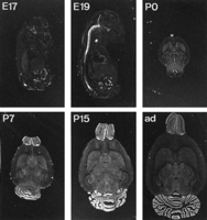
Developmental expression of NBC in rat brain.In situ hybridization was performed on frozen sections from rat brains of the indicated ages. Control sections hybridized with probe to which a 200-fold excess of unlabeled probe was added showed essentially no signal (data not shown). ad,Adult.

Cellular resolution of hybridization signal. Emulsion-dipped sections from P15 rats were exposed for 6 weeks and then developed and counterstained. Regions from the hippocampus (A, C) and the cerebellum (B, D) are shown at two magnifications. Dark-field photomicrographs show the hybridization signal as whitedots(A, B), whereas bright field shows the hybridization signal as darkdots (C, D). A, C, Labeling in the hippocampus reveals a lack of association of the hybridization grains with neurons. Arrowheads in C indicate clusters of grains between pyramidal neuronal cell bodies.B, D, Labeling in the cerebellum is consistent with expression in astrocytes and in particular in Bergman glia. Few grains are observed directly over the Purkinje cells indicated by arrowheads in D. The fields were photographed with a 10× and 40× objective using a Zeissaxioplan microscope. DG, Dentate gyrus;gr, granular layer; mol, molecular layer;p, Purkinje cell layer; so, stratum oriens; sp, stratum pyramidale; sr,stratum radiatum. E, F, To confirm the identity of the hybridizing cells, double labeling was performed using anti-GFAP antibody to identify astrocytes on the same section that was hybridized with a digoxigenin-labeled probe for NBC. An area from the cerebellum is shown; arrowheads indicate individual cells showing both cytoplasm immunoreactivity for GFAP (E) and a hybridization signal to the riboprobe for NBC (F). GL, granular layer;ML, molecular layer; PL, Purkinje layer. Similar sections stained only for GFAP showed the same pattern of staining (data not shown).
Overexpression of NBC in 3T3 cells
The mouse fibroblast cell line 3T3 does not normally express much if any NBC, as shown by Western blots (Fig.6A). We used this cell line to test the effect of overexpressing NBC on acid vulnerability. Either NBC or β-galactosidase as a control was stably expressed in 3T3 cells using retroviral vectors. Sister cultures were exposed to pH 6.8 for 24 hr. As seen in Figure 6, both sham-transfected and β-galactosidase-expressing 3T3 cells showed 15–20% cell death after 24 hr in BSS5.5 at pH 6.8 in the presence of both extracellular bicarbonate and sodium, whereas ∼40% of the NBC-overexpressing cells died, a significant increase in injury (Fig.6B). Both removing bicarbonate and adding DIDS reduced the injury to background levels. An immunoblot using a polyclonal antibody to NBC shows the level of NBC expression obtained by retroviral transfection of 3T3 cells compared with controls (Fig.6A). Morphological evidence of the difference in survival after exposure to pH 6.8 was obtained by trypan blue staining (data not shown) and was in agreement with the levels of LDH release.

A, Little NBC is detected by immunoblot in 3T3 cells (CTRL), whereas retroviral expression of NBC induces a large increase in the immunoreactive band for NBC. Purified brain plasma membrane (PM) is shown in the firstlane as a positive control, followed by NBC-expressing 3T3 cells (NBC), β-galactosidase-expressing 3T3 cells (lacZ), and control 3T3 cells. Equal amounts of protein were loaded.B, Overexpression of NBC renders 3T3 cells vulnerable to acid injury. Untransfected and control β-galactosidase-expressing 3T3 cultures exposed to BSS5.5 at pH 6.8 for 24 hr showed little injury, whereas 3T3 cells overexpressing NBC suffered ∼40% cell death. Substituting for bicarbonate or adding 1 mmDIDS significantly reduced the injury of NBC-overexpressing cells. * indicates significant difference from control and lacZ;p < 0.05; n = 10–16.
DISCUSSION
We have isolated a functional cDNA clone of the electrogenic sodium bicarbonate cotransporter from rat brain. This sequence is the same NBC variant recently identified from pancreas and differs in the 5′-untranslated and N-terminal coding sequence from the NBC cDNA characterized previously from kidney. The transporter is electrogenic, requires sodium and bicarbonate, and is inhibited by DIDS, thus showing the principal characteristics of NBC. Extracellular pH 6.8 injury of primary cultured astrocytes requires extracellular sodium and bicarbonate and is also inhibited by DIDS. The association of NBC with acid sensitivity is underlined by the ability to confer acid sensitivity on 3T3 cells by overexpressing NBC.
The immediate response of NBC to lowering extracellular bicarbonate and pH is to run in the outward direction (Munsch and Deitmer, 1994) acidifying intracellular pH, but there were no previous studies on the function of the transporter in the setting of prolonged reduction of extracellular bicarbonate at pH 6.8, as studied here. We demonstrate that the intracellular pH measured with BCECF in the presence of extracellular bicarbonate is higher than that in its absence, after 3 hr at extracellular pH 6.8. This is consistent with the transporter running in the inward direction after equilibration to the new buffer condition. The severity of intracellular acidosis did not correlate with injury. Protection of astrocytes from ischemia-like injury by DIDS is thus reminiscent of the response of myocardial cells. Inhibition of intracellular alkalinization by inhibition either of the sodium hydrogen exchanger or of bicarbonate transport has been shown to be highly protective in the setting of myocardial ischemia (Karmazyn, 1998). In the case of myocardial ischemia too rapid alkalinization of intracellular pH is associated with a worse outcome.
Protection by reducing extracellular sodium may at first be surprising, because a variety of transporters depend on the sodium gradient for their normal function. However, sodium overload has been shown to contribute to anoxic neuronal injury, and substitution for Na+ with an impermeant ion was protective (Friedman and Haddad, 1994a). Elevation of intracellular Na+ is thought to contribute to reversal of Na+/Ca2+exchange and to worsen calcium overload. Although the mechanism of astrocyte injury induced by lowering medium pH to 6.8 is not known, it is possible that sodium overload contributes to the injury. Prolonged sodium influx could contribute to calcium overload by reversal of Na+/Ca2+exchange and contribute to energy depletion by chronic activation of the Na+/K+ATPase. The Na+/K+ATPase has been reported to saturate above intracellular Na+ concentrations of 30 mm(Collins et al., 1992), and intracellular Na+ concentrations above 50 mmhave been reported in anoxic neurons in vitro (Friedman and Haddad, 1994b).
While this work was in progress, the N-terminal NBC variant identified by us in brain was reported from pancreas (Abuladze et al., 1998). The Northern analysis presented by Abuladze et al. (1998) and our RT-PCR data agree in finding this splice variant in many tissues, whereas the kidney splice variant is restricted primarily to kidney. Two variants with different N-terminal sequences allow for differential functional regulation, possibly by phosphorylation (Abuladze et al., 1998). The different N-terminal sequences in the two NBC forms may also allow for association with different intracellular or membrane proteins. In the case of the anion exchanger AE1, sorting to the plasma membrane or retention in intracellular membranes is determined by the N-terminal sequence (Cox et al., 1995). Finally, the N-terminal sequences could determine the formation of heteromeric transporters, if indeed the transporter is a functional multimer.
Physiological studies of NBC in different cell types and species have demonstrated that it can transport either two or three bicarbonate ions per sodium ion. It is intriguing to ask whether the different ratios observed physiologically reflect expression of different genes or regulation of a single transporter. The existence of the NBC variants suggests the possibility of differences in function being specified at the level of promoter choice or splice variation. Further studies and possible identification of additional bicarbonate transporters will be needed to answer this question.
In situ hybridization shows that the transporter is expressed throughout the brain, beginning at approximately the time of birth and persisting throughout adulthood. The primarily late developmental expression suggests that this transporter is critical during the later stages of brain development. It is expressed during the time of generation and maturation of astrocytes. Appropriate regulation of both intracellular and extracellular pH may be increasingly important with brain maturation, accounting for the time course of expression of this protein. That it is widely distributed throughout the brain suggests it serves a basic function in all brain regions. It is likely that proper functioning of the pH regulatory NBC transporter is important to proper neuronal function, although the transporter appears to be present primarily in glial cells. The expression of NBC may contribute to the different patterns of injury caused by ischemia in the perinatal period compared with the adult. Astrocyte development in the brain may also impact the outcome from ischemia.
Astrocytes are now known to perform many essential functions including modulating neuronal excitability (Ransom, 1992), regulating the extracellular concentrations of ions (Newman, 1995) and neurotransmitters (Schousboe and Westergaard, 1995), providing a critical metabolic link between the blood supply and neurons (Tsacopoulos and Magistretti, 1996), and protecting neurons from excitotoxic (Rosenberg and Aizenman, 1989; Dugan et al., 1995) and oxidative injury (Raps et al., 1989; Sagara et al., 1993; Desagher et al., 1996). Thus neuronal function and survival are inextricably linked to glial function and survival. Preventing glial impairment is highly likely to contribute to improved neuronal survival during in vivo ischemia. The identification of the sodium/bicarbonate cotransporter as participating in astrocyte injury provides a novel target for brain protective strategies. This is the first glial-specific mechanism of injury to be identified. To test directly the role of NBC on the outcome from cerebral ischemia will require changing the level of NBC expression in astrocytes in vivo. Much as the idea of excitotoxicity suggested a variety of strategies to block neuron-specific injury, it may now be possible to target glial-specific injury mechanisms.
Footnotes
- Received August 16, 1999.
- Revision received October 18, 1999.
- Accepted November 4, 1999.
-
This work was supported in part by Boehringer Ingelheim and the Schilling Foundation (H.M.), sabbatical leave (R.G.G.), National Institutes of Health Grant GM 49831 (R.G.G.), and a NATO Science Fellowship (J.A.v.H.). We thank Ulla Amtmann for expert technical assistance, Walter Boron for the anti-NBC antibody, and Peter Seeburg for helpful discussions. The GenBank accession number for NBC isAF210250.
Correspondence should be addressed to Dr. Rona Giffard, Department of Anesthesia, S272, Stanford University School of Medicine, Stanford, CA 94305-5117. E-mail: rona.giffard{at}stanford.edu.
Dr. van Hooft's present address: Institute of Neurobiology, University of Amsterdam, Kruislaan 320, NL-1098 SM Amsterdam, The Netherlands.
Dr. Papadopoulos's present address: Neurosurgery, Atkinson Morley's Hospital, Wimbledon, London SW20 0NE, United Kingdom.
- Copyright © 2000 Society for Neuroscience




