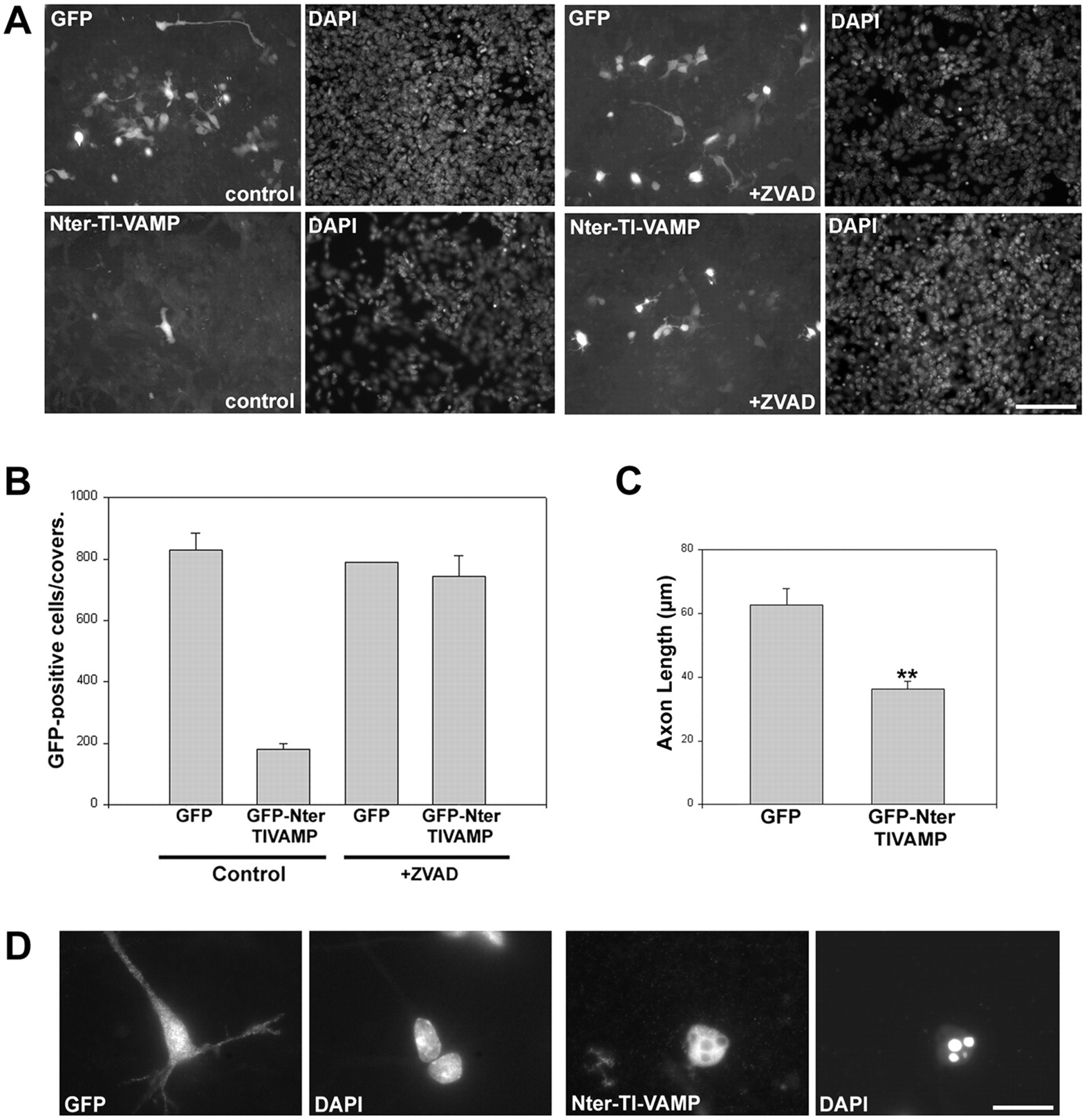
Expression of the N-terminal domain of TI-VAMP induces apoptosis. A, Corticostriatal neurons from intact embryonic brains were electroporated with the indicated constructs and cultured for 24 hr in the absence (left panels) or presence (right panels) of the caspase inhibitor zVAD. Observe the increase in the number of transfected cells in zVAD-treated Nter-TI-VAMP-electroporated cells compared with nontreated cells. In the case of GFP-electroporated cells, there is no difference between zVAD-treated or nontreated cells. Scale bar, 100 μm. B, Quantification of the apoptotic effect of the N-terminal domain of TI-VAMP in cells treated as inA; shown are the mean values (±SEM) of the number of positive cells on each coverslip. C, Quantification of the effect in axonal length of the expression of the N-terminal domain of TI-VAMP in cells treated as in A. Shown are the mean values (±SEM) of a minimum of 40 cells. D, Neurons infected with Aav carrying GFP or Aav carrying GFP-Nter-TI-VAMP fixed 3 d after infection. A representative cell of each type is shown. Note that the cell expressing GFP displays neurites and a normal nucleus compared with a noninfected cell, whereas the cell expressing Nter-TI-VAMP is round, with no neurites and presents a typical apoptotic nucleus as seen with DAPI staining. Scale bar, 20 μm.


