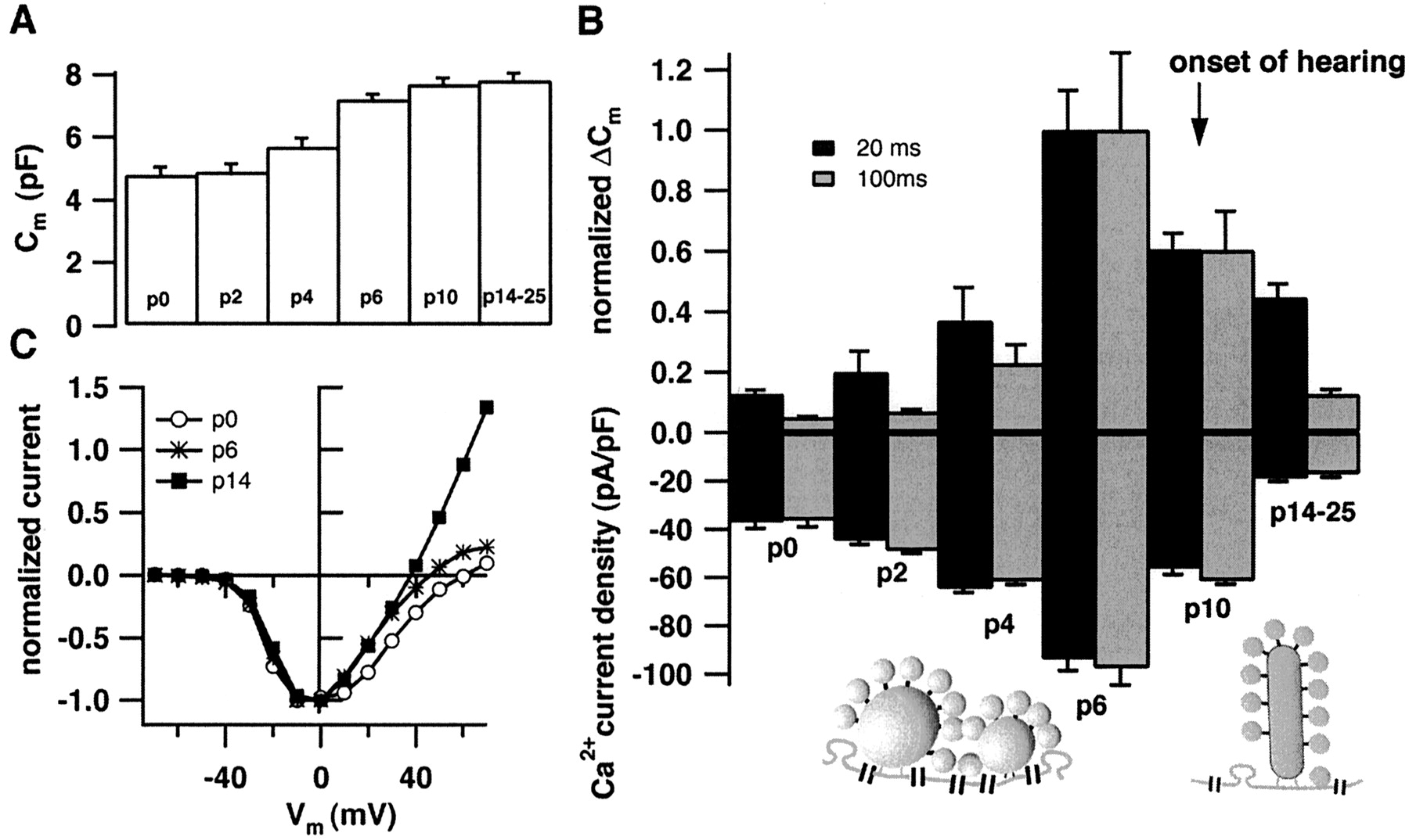
Developmental changes in Ca2+current and exocytosis. A demonstrates the increase in basal cell capacitance during the course of maturation (P0,n = 13; P2, n = 2; P4,n = 10; P6, n = 10; P10,n = 9; and P14–P25, n = 28).B displays normalized ΔCm-responses (top panel) to 20 or 100 msec long depolarizations to −5 mV (number of cells as in A, normalized to the average responses of P6 cells to either depolarization). The bottom panel shows the Ca2+ current densities for 20 and 100 msec depolarizations (peak Ca2+ currents normalized to the cell capacitance). The arrow indicates the onset of hearing. The diagrams depict the preferential abundance of multiple spherical bodies at the active zones of immature IHCs and the typical presence of only one plate-like ribbon at the mature afferent synapse. C displays representative current voltage relationships for three different developmental stages after subtracting the leak current (estimated by a linear fit to the hyperpolarized portion of the data).


