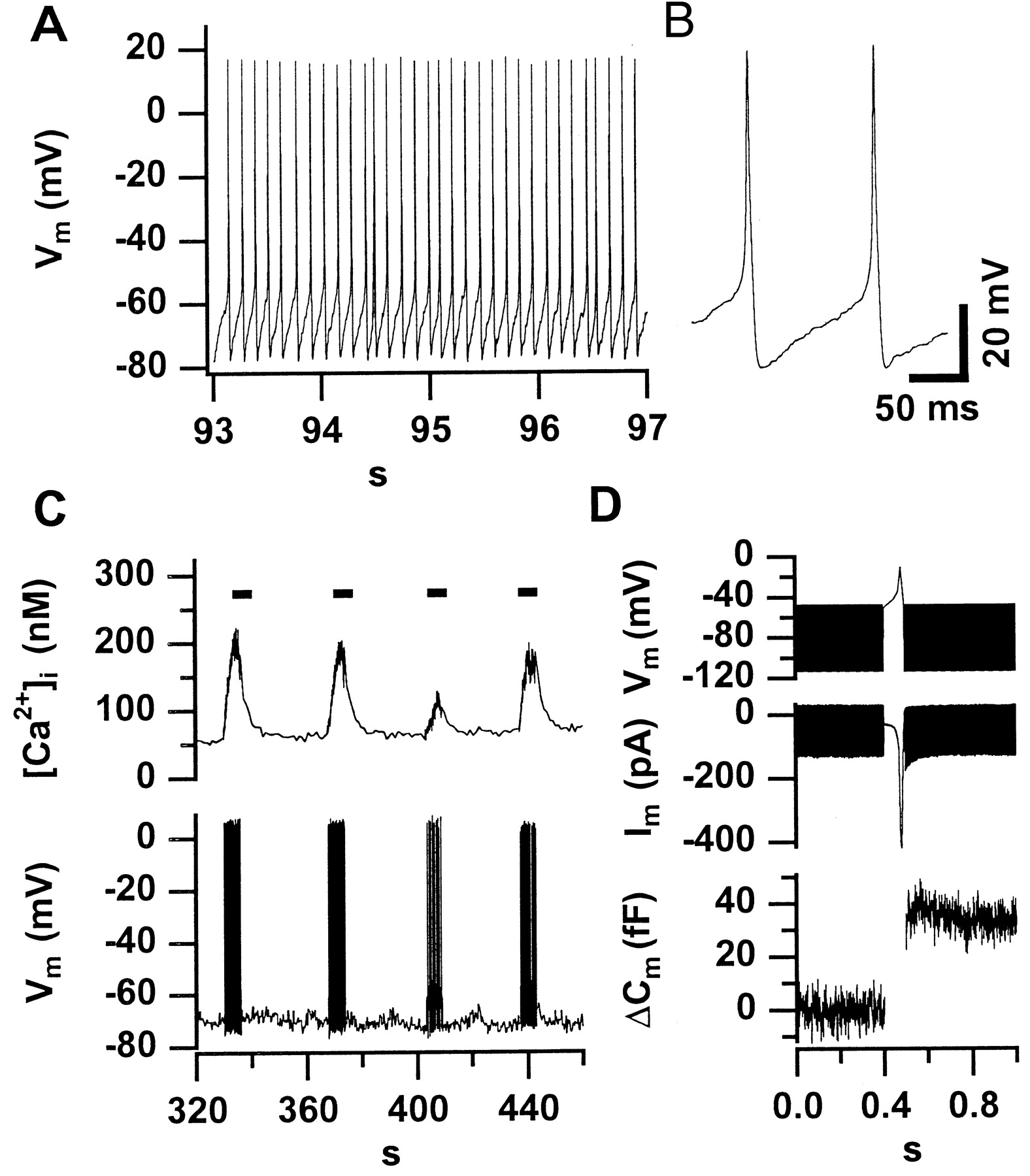
Spontaneous action potentials trigger exocytosis and increase [Ca2+]i in immature IHCs.A displays an AP train recorded in the whole-cell configuration in the absence of any holding current from a representative P6 IHC. In this particular cell, spontaneous activity was observed during the first 260 sec of the whole-cell recording.B displays two spontaneous APs from the same cell at higher time resolution. C shows a low-time resolution plot of [Ca2+]i and membrane potential (Vm) of an IHC in which we applied 10 pA of depolarizing current for short intervals (indicated bybars) after spontaneous activity had ceased.D shows an average of three secretory responses of two IHCs from a P6 mouse to a single action potential. We used the voltage-clamp mode to measure capacitance before and after an AP-like voltage-waveform to −10 mV. The time periods ofCm measurements can be recognized from the band-like (sinusoidal) signals in the voltage and currenttraces.


