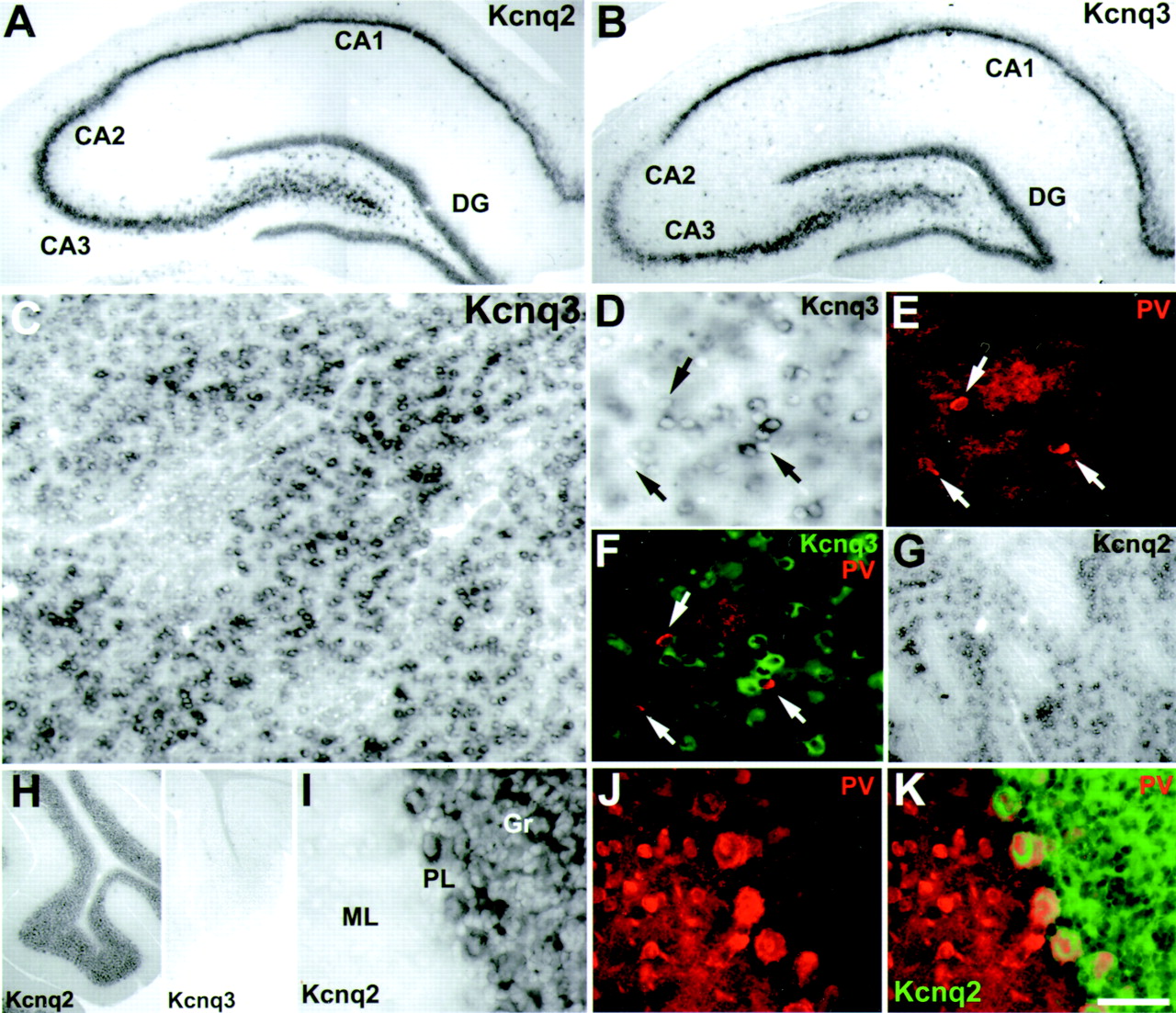
Kcnq2 and Kcnq3 mRNA transcripts overlap in the hippocampus and caudate/putamen, but not the cerebellum.A–B, Comparison of Kcnq2 and Kcnq3 transcripts in the hippocampus. A, Abundant Kcnq2 expression was detected in CA1–CA3 pyramidal cells and granule cells of the DG.B, Strong Kcnq3 expression was found in similar neurons but was weaker than Kcnq2 in CA2. C–G, Kcnq3 and Kcnq2 expression in the caudate/putamen (CPu).C, Low-magnification bright-field image showing strong Kcnq3 expression in the large majority of CPu neurons.D–F, Kcnq3 is not expressed in PV-immunoreactive neurons in the CPu. D, High-magnification bright-field image of Kcnq3-positive neurons in the CPu. E, Immunofluorescent detection of PV-containing neurons in same section asD. F, Overlay of D and Ewith Kcnq3 pseudocolored green. Note that no PV neurons were Kcnq3 positive (arrows). G, Kcnq2 is also located in the caudate/putamen. H–K, Kcnq2, but not Kcnq3, is expressed in the cerebellar cortex. H, Low-magnification image of Kcnq2 (left) and Kcnq3 (right) expression in the cerebellar cortex.I–K, Kcnq2 is located in the granule and Purkinje cell layer of the cerebellum. I, High-power bright-field image showing Kcnq2 expression in the cells of the Purkinje layer (PL) and granule layer (Gr) but not the molecular layer (ML). J, Same image asI, with immunodetection of PV-reactive Purkinje cells and interneurons of the molecular layer. K, Overlay of I and J, with Kcnq2 pseudocoloredgreen. Scale bar (shown in K):A, B, 275 μm; C, 250 μm; D–F, 100 μm;G, 300 μm; H, 475 μm;I–K, 50 μm.


