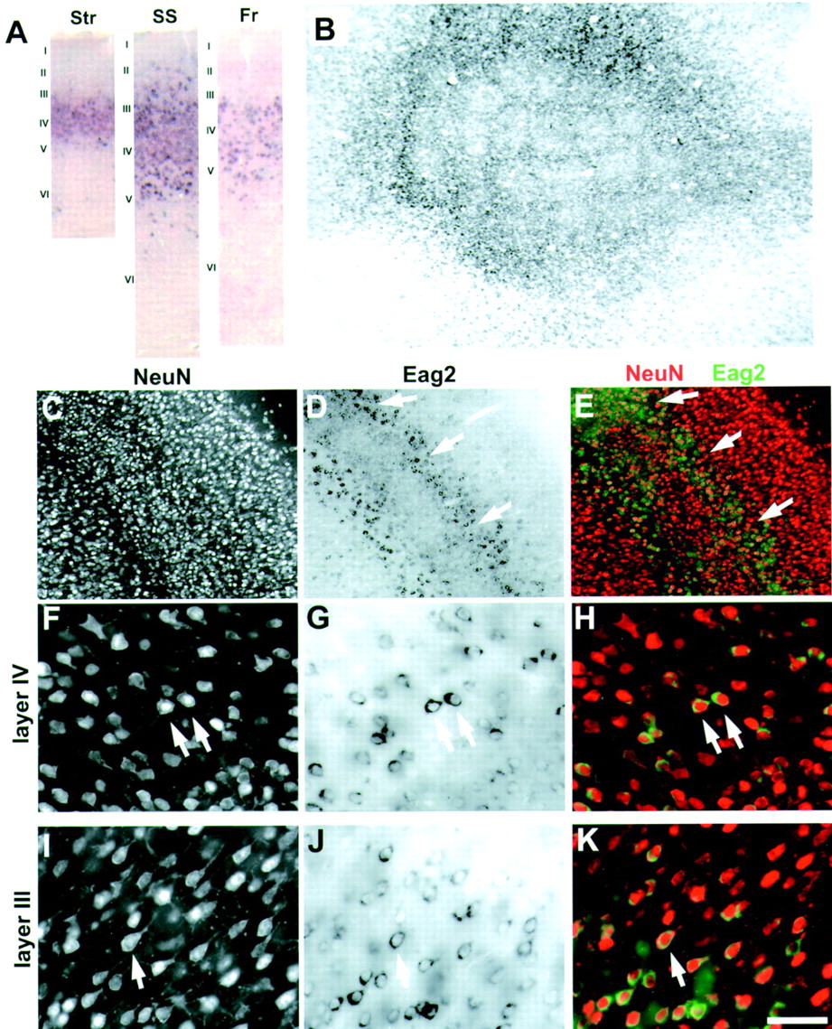
Characterization of eag2 expression in the cerebral cortex. A, Changes in eag2 expression with cortical region. Eag2-expressing neurons are more abundant in somatosensory cortex (SS) as compared with the striate (Str) and frontal (Fr) cortical regions.B, ISH for eag2 in tangential sections through rat somatosensory barrel cortex reveals a whisker barrel pattern with hollow centers. C–E, Combined ISH for eag2 and immunofluorescent detection of NeuN in a coronal section through rat somatosensory barrel cortex. C, Fluorescent detection of all neurons using NeuN antibodies. D, Same section in bright field showing labeling for eag2 by ISH. Note that eag2 staining demarcated cortical layer IV with strong labeling of neurons lining the barrel sides (arrows), as well as neurons on the margins between layer IV and neighboring layers. E, Overlay ofC and D with eag2 pseudocoloredgreen, and NeuN red. F–K, High-magnification images of C identifying eag2-positive neurons along barrel sides in cortical layers IV (F–H) and in deep cortical layer III (I–K). Note that eag2-positive neurons in layer IV are small and non-pyramidal and have a star-shaped appearance (F–H, arrows). In contrast, eag2-positive cells in deep layer III are clearly pyramidal in shape with identifiable apical dendrites that are orientated toward the pia surface (I–K,arrow). Scale bar (shown in K):A, 400 μm; B, 575 μm; C–E, 500 μm;F–K, 50 μm.


