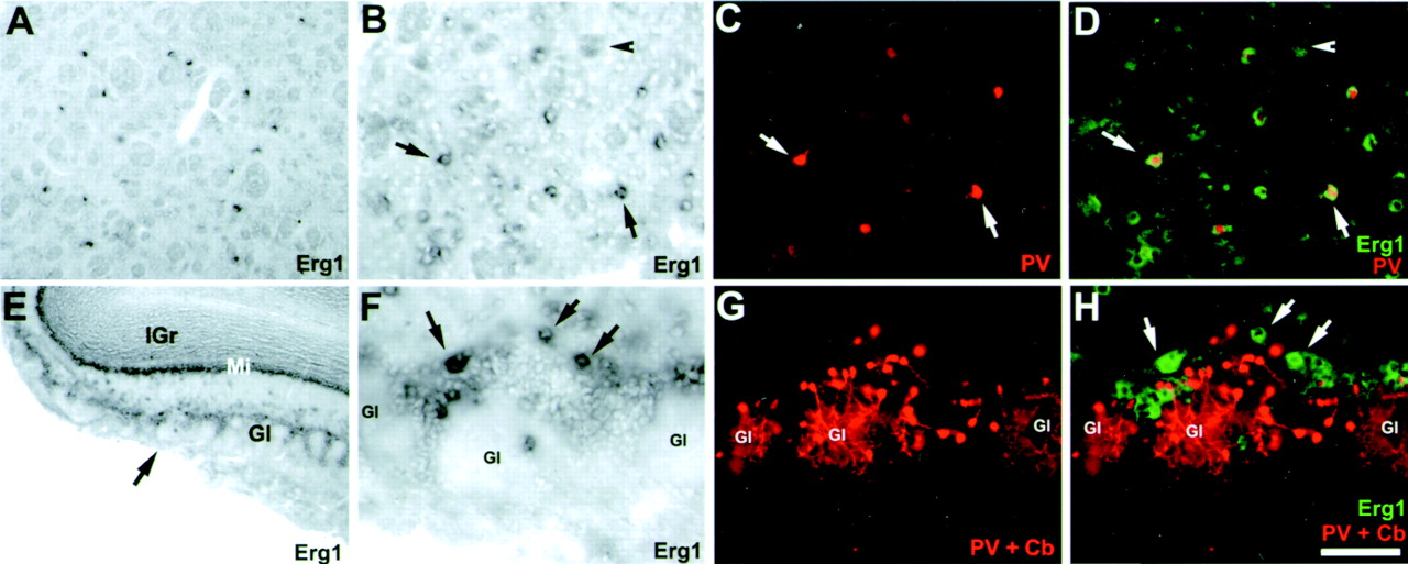
Erg1 transcripts are located in a few scattered PV-positive cells in the caudate/putamen but do not colocalize with PV or Cb in the olfactory bulb. A–D, Erg1 transcripts are located in PV-positive interneurons of the caudate/putamen (CPu). A, Low-magnification bright-field image of erg1-expressing cells in the CPu. Note that cells are strongly labeled but scattered and few in number. B, Higher magnification image of erg1-positive neurons in the CPu. Fiber tracts appear as light gray (arrowhead).C, Immunofluorescent detection of PV-containing neurons.D, Overlay of B and C with erg1 signals pseudocolored green. Note that most PV-positive neurons also express erg1 (B–D,arrows). E–H, Erg1-expressing neurons in the periglomerular layer of the olfactory bulb are not immunoreactive for PV or Cb. E, Low-magnification bright-field image of erg1 in the olfactory bulb. F, High magnification of E (arrow) showing three glomeruli (Gl). Note that erg1 expression is strong and located in large neurons within the periglomerular layer (arrows). G, Immunofluorescent detection of PV- and Cb-positive periglomerular neurons (PV and Cb monoclonal antibodies were mixed and detected with the same secondary antibody).H, Overlay of F and G with erg1 labeling pseudocolored green. Note that none of the large erg1-positive neurons are PV or Cb positive. Scale bar (shown inH): A, 250 μm; E, 500 μm; B–D, F–H, 100 μm.


