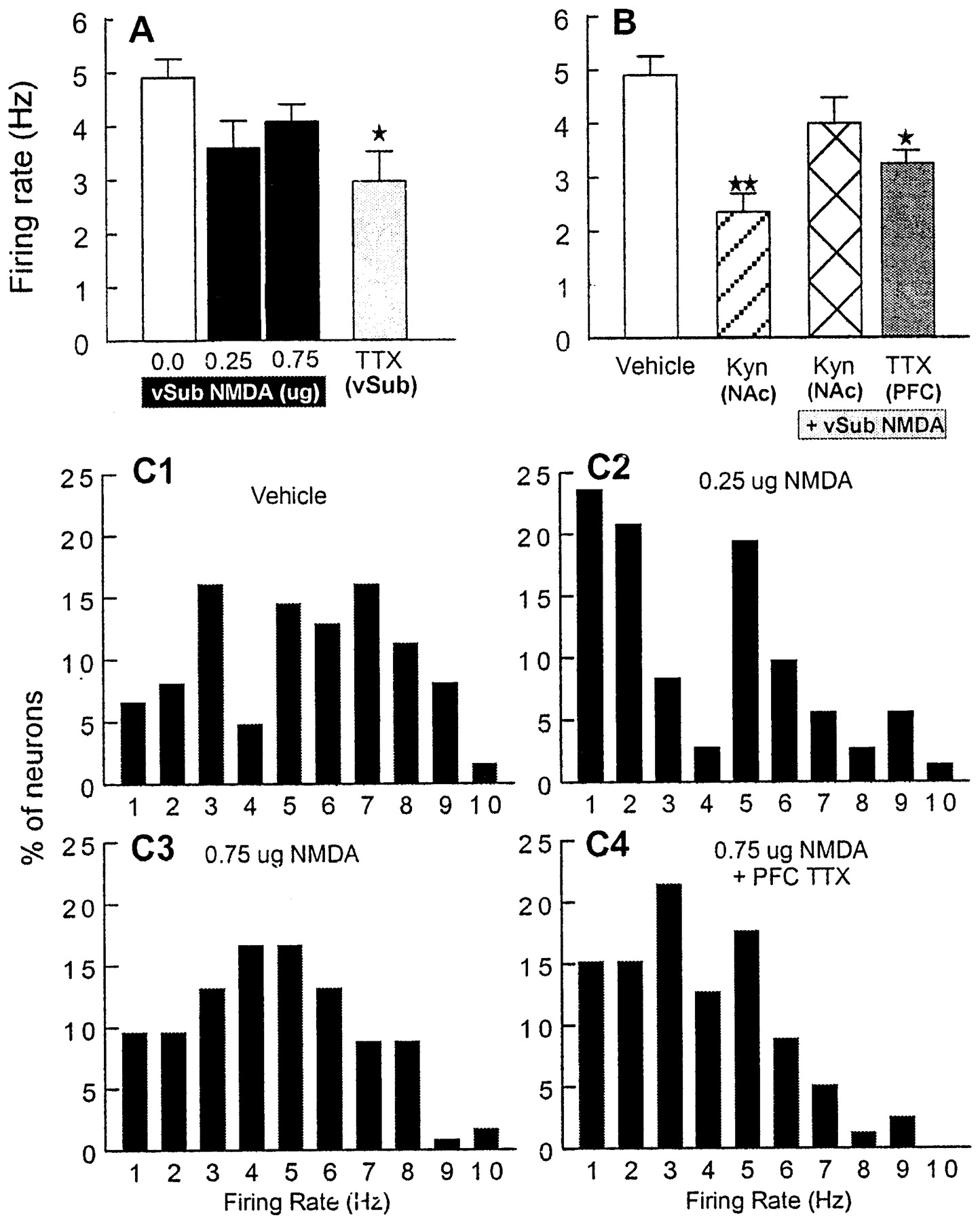
Modulation of DA neuron firing rate by the vSub, the NAc, and the mPFC. A, In contrast to the effects on spontaneous activity, activation of the vSub did not change significantly the VTA DA neuron firing rate. Nonetheless, inactivation of the vSub did decrease VTA DA cell firing. The mean firing rate of DA neurons (+SEM) recorded from rats receiving vehicle infusions (white bar), 0.25 or 0.75 μg of NMDA (black bars), or TTX (gray bar) into the vSub is shown. B, Blockade of glutamate transmission in the NAc attenuates the DA neuron firing rate. The mean firing rate of DA neurons (+SEM) recorded from rats receiving vehicle infusions (same asA; white bar), infusions of kynurenic acid into the NAc in combination with vehicle infusions into the vSub (hatched bar), infusions of kynurenic acid into the NAc in combination with 0.75 μg of NMDA into the vSub (cross-hatched bar), or infusions of TTX into the mPFC in combination with 0.75 μg of NMDA into the vSub (gray bar) is shown. *p < 0.05 and **p < 0.01 versus control. C, Distribution of mean firing rates of all DA neurons recorded from rats receiving vehicle injection (C1), 0.25 μg of NMDA (C2) or 0.75 μg of NMDA (C3) into the vSub, or infusions of TTX into the mPFC in combination with 0.75 μg of NMDA into the vSub (C4) is shown. This suggests that the increase in DA cell spontaneous activity after vSub stimulation may be caused by the activation of slow-firing DA neurons.


