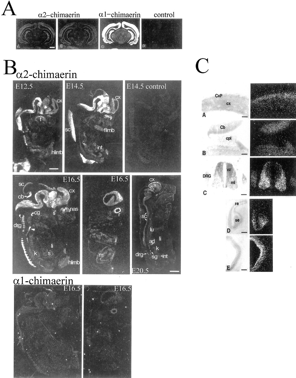
α2-chimaerin mRNA, but not α1-chimaerin mRNA, is highly expressed in embryonic nervous system. A,Specificity of detection of α2- and α1-chimaerin mRNA was determined by in situ hybridization of coronal sections of adult rat brain with oligonucleotide probes complementary to α2-chimaerin N-terminal coding sequence (AA ), an α2-chimaerin oligonucleotide spanning the point of sequence divergence from α1-chimaerin (AB ), α1-chimaerin 5′sequence, showing the higher level of expression of α1-chimaerin in adult brain (AC ), and an α2-chimaerin sense strand control oligonucleotide (AD ). Scale bar, 2 mm.B, α2-chimaerin mRNA expression during embryonic development. At embryonic day 12.5 (E12.5), high levels of specific labeling were detected in central and peripheral nervous system with most intense signal in caudal structures. E14.5 and E16.5 embryos show progressively more rostral hybridization; the signal could be competed by a 20-fold excess concentration of unlabeled oligonucleotide (E14.5 control); E16.5 embryos show strong hybridization to cerebral cortex and in a more lateral E16.5 section hybridization to the retina (r). Although the nervous system clearly exhibits the highest levels of hybridization throughout embryonic development, various other structures including limb buds, smooth and striated muscle, and developing organs such as the kidney and lung are also slightly labeled at longer exposures. At 20 d (E20.5) moderate levels of hybridization are detected in most neural tissues with higher levels in sympathetic ganglia and in a band outlining the cerebral cortex.ad, Adrenal gland; cb, cerebellum;cg, cervical ganglion; col, colliculus;cx, cortex; drg, dorsal root ganglion;int, intestine; flimb, forelimb;hlimb, hindlimb; k, kidney;li, liver; lu, lung; m, muscle; nas, nasal epithelium; r, retina;s, stomach; sc, spinal cord;sg, sympathetic ganglion; t, tongue;5, trigeminal ganglion. Hybridization to α1-chimaerin-specific oligonucleotide was detectable at E16.5. Central and lateral sections show very low levels of expression [compare with hybridization to α2-chimaerin specific oligonucleotide in corresponding (E16.5) sections above]. Scale bars: E12.5–E16.5, 1.5 mm; E20.5, 3.5 mm. C, α2-chimaerin mRNA expression in embryonic rat tissues at E12.5 and E16.5. Light-field photomicrographs of counterstained sections and corresponding dark-field in situ hybridization in emulsion-dipped slides: (CA ) E16.5 cerebral cortex. Note the higher expression in the cortical plate (CxP) relative to cortical neuroepithelium (cx). CB , E16.5; there is expression in the cerebellar anlage (Cb) but not in the adjacent choroid plexus (cpl).CC , E12.5; α2-chimaerin mRNA is present in dorsal root ganglion (DRG) and at high levels in spinal cord mantle layer (ml) but not in the ventricular zone (vz). CD , E16.5 olfactory sensory (se) but not respiratory epithelium (re) shows hybridization.CE , E16.5; labeling of the retina is most intense over the inner band of cells. Scale bar, 150 μm.


