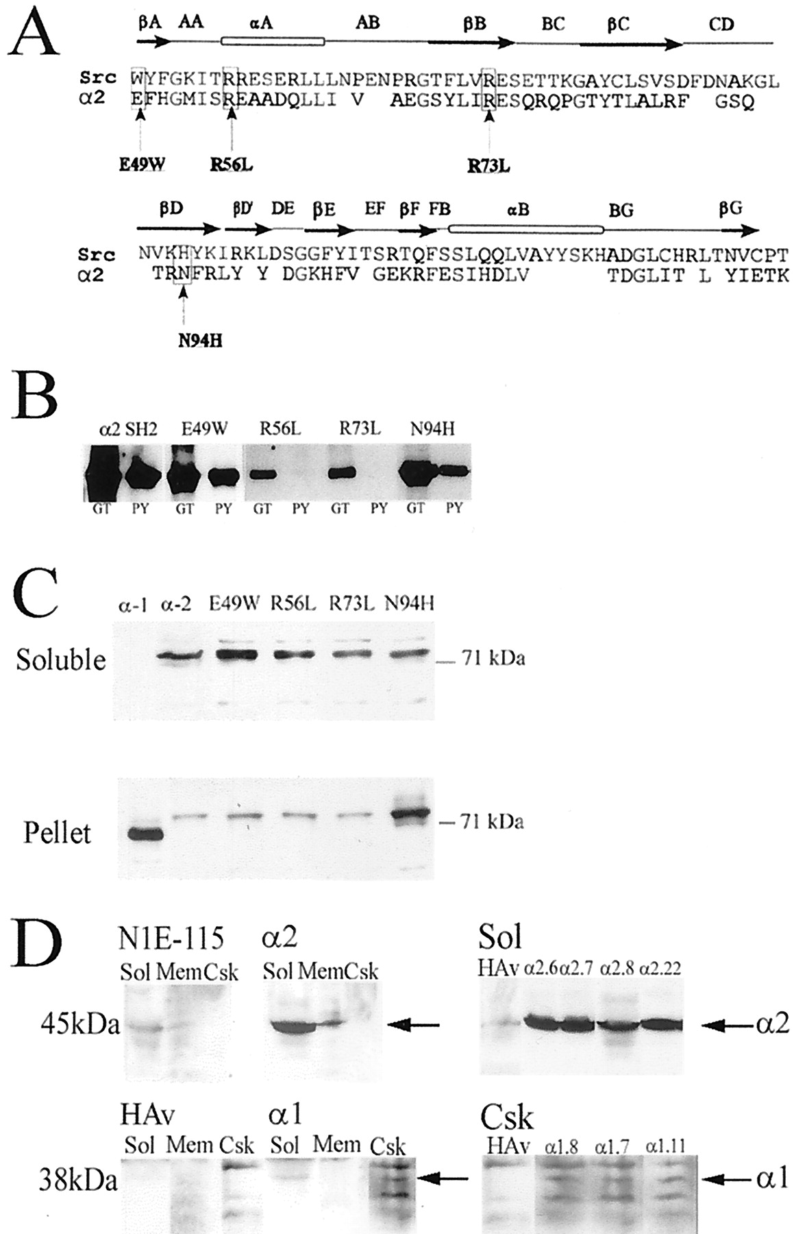
α2-chimaerin SH2 domain mutations and their effect on protein distribution in transfected cells. A,Alignment of chimaerin SH2 domain with Src SH2 domain. Notation above the alignment refers to SH2 domain secondary structure (Waksman et al., 1993). Mutations were made at positions equivalent to Src βA1, αA2, βB5, and βD4, residues likely to be involved in phosphopeptide interaction. B, Effect of SH2 domain mutations on binding to phosphotyrosine-agarose. Chimaerin SH2 domain and four SH2 domain mutants, E49W, R56L, R73L, and N94H, expressed as GST fusion proteins were bound to either glutathione-agarose (GT) or phosphotyrosine-agarose (PY) columns eluted in SDS Laemmli buffer and analyzed by Western immunoblotting with α2-chimaerin antibody. C, Distribution of chimaerin isoforms and SH2 domain mutants. COS-7 cells were transiently transfected with GFP/α1-chimaerin, GFP/α2-chimaerin, GFP/α2-chimaerin E49W, GFP/α2-chimaerin R56L, GFP/α2-chimaerin R73L, or GFP/α2-chimaerin N94H. Soluble and pellet fractions were analyzed by Western immunoblotting with anti-GFP monoclonal antibody (Clontech). Similar results were obtained in experiments with transiently transfected HA-tagged constructs in place of GFP.D, Permanent cell lines overexpressing α2-chimaerin or α1-chimaerin were fractionated into soluble (Sol), solubilized membrane (Mem), and cytoskeletal (Csk) fractions. They were subject to Western immunoblotting using anti α2-chimaerin polyclonal antibodies to detect α2-chimaerin or anti-HA monoclonal antibody for α1-chimaerin. Soluble fractions from a series of α2-chimaerin lines (Sol) and cytoskeletal fractions from a series of α1-chimaerin lines (Csk) are also shown. The positions of α2- (45 kDa) and α1-chimaerin (38 kDa) are indicated (arrows). Fractions from parent N1E115 cells and empty vector containing cells (HAv) are also shown.


