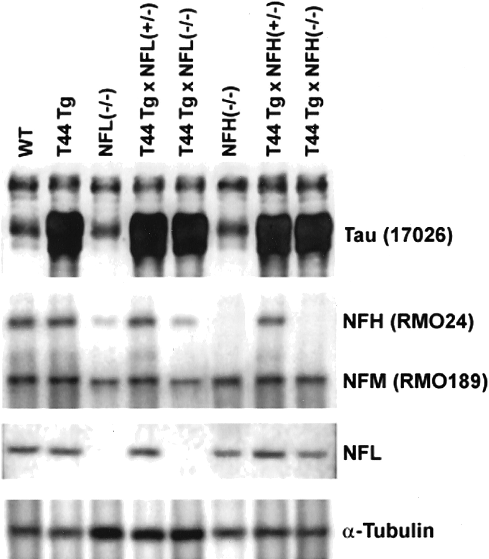
Analysis of protein expression in cortices. Western blot analysis of tau, NFs, and α-tubulin isolated from cortices of the different lines of mice. Six-month-old mice from each group were used. 17026, an anti-recombinant tau antibody that recognizes both human and mouse tau, shows the expression level of tau in each line of mice. Endogenous mouse tau protein levels are comparable in WT, NFL−/−, and NFH−/− mice, and overexpressed human tau protein levels in T44 Tg, T44 Tg;NFL+/−, T44;NFL−/−, T44;NFH+/−, and T44;NFH−/− mice were ∼10-fold higher than endogenous mouse tau. NFH and NFM protein levels were decreased ∼85 and 50%, respectively, in the cortices of NFL−/− and T44 Tg;NFL−/− mice, whereas the level of α-tubulin was dramatically increased compared with WT mice. NFL protein levels were decreased ∼30% in NFH−/− and T44 Tg;NFH−/− mice without a change in NFM protein levels. Equal amounts (10 μg for tau; 20 μg for NFs and α-tubulin) of mouse cortical samples were loaded on each gel lane.


