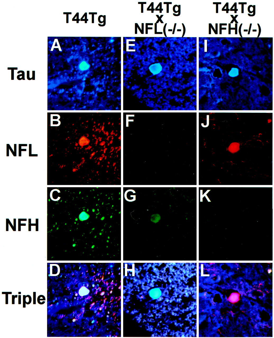
Fig. 4.
Triple immunofluorescence staining of spheroids in the spinal cord of 6-month-old T44, 15-month-old T44;NFL−/−, and 6-month-old T44 Tg;NFH−/− mice. The sections were stained with the T14 mouse anti-human tau mAb (A, E,I), the rabbit anti-NFL antibody (B, F, J), and the DP1.6 rat anti-NFH mAb (C, G,K). A triple fluorescent filter cube was used to visualize colocalization of tau, NFL, and NFH (D,H, L). All photomicrographs are at the same magnification. Scale bar, 10 μm.


