In the article “The OMP–lacZ Transgene Mimics the Unusual Expression Pattern ofOR-Z6, a New Odorant Receptor Gene on Mouse Chromosome 6: Implication for Locus-Dependent Gene Expression,” by Martina Pyrski, Zheng Xu, Eric Walters, Debra J. Gilbert, Nancy A. Jenkins, Neal G. Copeland, and Frank L. Margolis, which appeared on pages 4637–4648 of the July 1, 2001 issue, Figure 6 was incorrectly printed as a black and white figure. The correct color version of the figure, as well as the legend, is printed here.
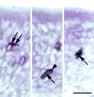
The three bright-field images illustrate the criteria by which olfactory neurons coexpressing thelacZ transgene and OR-Z6 were identified.OR-Z6 in situ hybridization was performed using a digoxigenin-labeled OR-Z6 riboprobe, and hybridization signals were detected and visualized using an alkaline phosphatase-conjugated anti-digoxigenin antibody and a substrate that developed a purple precipitate. Left, Hybridization signals were most prominent in the apical cytoplasmic portion and appeared triangular (arrows). In addition we observed a thin line surrounding the nucleus; the nucleus itself was usually white. Right, In contrast, ORNs that solely express the lacZ transgene (arrow) were gray, including the cytoplasm covering the nucleus. Expression of β-galactosidase was detected using a primary anti-β-galactosidase antibody and visualized using a substrate yielding a gray precipitate.Middle, The double-labeled neuron shows a combination of both features described. The gray cytoplasm covering the nucleus indicates β-galactosidase expression (arrow), whereas the peak-shaped purple precipitate in the apical cytoplasm corresponds to OR-Z6 in situ hybridization (arrowhead). The three differently labeled neurons shown derive from the same coronal cryosection of a 35-d-old H-lacZ6 mouse and were closely associated on endoturbinate-II. Scale bar, 20 μm.
- Copyright © 2001 Society for Neuroscience




