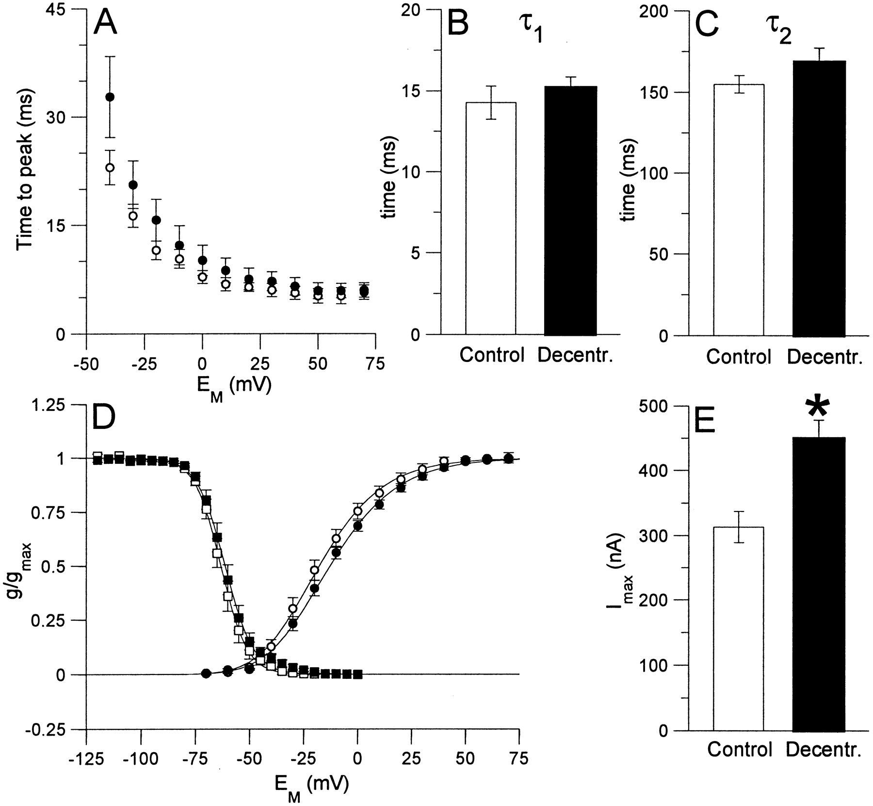
Effect of decentralization on parameters of IA in PD neurons after 6 d in organ culture. All values are means ± SEM (control preparation,n = 7; decentralized preparations,n = 8). A, Time-to-the peak current as a function of membrane potential for activation ofIA in PD neurons under control conditions (open circles) and in decentralized preparations (filled circles). The time-to-peak current did not differ significantly between the two conditions. B,C, During a maintained voltage pulse,IA decays with two time constants, τ1 (fast) and τ2 (slow). Both time constants did not differ significantly between control conditions and decentralized preparations, as demonstrated for τ1(B) and τ2(C) for a voltage pulse to +50 mV.D, Conductance–voltage curves for activation (circles) and inactivation (squares) ofIA under control conditions (open symbols) and in decentralized preparations (filled symbols). Values are calculated as a fraction of the maximal conductance under control conditions in each experiment. The activation curves were generated from peak currents after a maximally de-inactivating prestep to −120 mV. The curves are fits to a third-order Boltzmann relation (Eq. 1, Materials and Methods) with the following parameters: control,Vact = −41.7 mV, s= −17.7 mV; decentralized, Vact = −38.2 mV, s = −18.9 mV. The curves for steady-state inactivation were generated from peak currents to voltage pulses to +50 mV after the neurons were held at the indicated potential (prepulse) for 1.5 sec. The curves are fits to a first-order Boltzmann relation (Eq. 1, Materials and Methods), with the following parameters: control, Vinact = −63.2 mV,s = 6.0 mV; decentralized,Vinact = −61.3 mV,s = 6.5 mV. E, Maximal current amplitude elicited by voltage pulses to +70 mV. After decentralization, the mean maximal current amplitude was significantly increased by 43.4%, from 313.6 ± 24.3 nA to 449.6 ± 27.7 nA (*p < 0.01).


