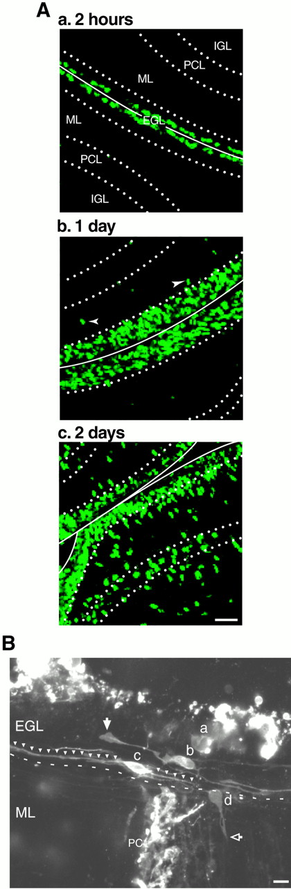
A, Time course of translocation of postmitotic granule cells in the EGL. P10 mice were injected with BrdU intraperitoneally and killed 2 hr (a), 1 d (b), and 2 d (c) later. Two hours after injection, BrdU-labeled cells were localized at the top level of the EGL. One day after injection, the BrdU-labeled cells occupied the entire EGL. Two days after injection, approximately half of BrdU-labeled cells left the EGL and translocated to the ML, PCL, and IGL. B, Morphology of granule cell precursors and postmitotic granule cells in the EGL of P10 mouse cerebellum. The granule cells in the EGL at the pyramis were visualized 2 hr after DiI staining. The granule cell precursor (a) had a round soma without any long processes. Postmitotic granule cells (b, c) had spindle-shaped cell bodies oriented parallel to the longitudinal axis of the folium with two horizontal processes. On the EGL–ML border, the postmitotic granule cell (d) had a rounded cell body with vertically oriented short processes penetrating the ML. Scale bars, 10 μm. PC, Purkinje cell.


