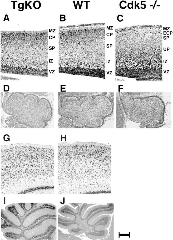
Histological analysis of TgKO mice brain compared with WT and Cdk5 −/− mouse brains from E18.5 embryos (A–F) and adult mice (G–J). A, The sagittal section of the cerebral cortex from the TgKO mouse embryo at E18.5. The thick cortical plate (CP) beneath the marginal zone (MZ) is clearly distinguished. Below the cortical plate, the subplate (SP) and the intermediate zone (IZ) are clearly recognized. B, The sagittal section of the cerebral cortex from the WT mouse embryo at E18.5. The cortex consists of the marginal zone (MZ), the cortical plate (CP), the subplate (SP), the intermediate zone (IZ), and the ventricular zone (VZ). C, The sagittal section of the cerebral cortex from the Cdk5 −/− mouse embryo at E18.5. The marginal zone (MZ) is followed by the early cortical plate (ECP), the subplate (SP), the underplate (UP), the intermediate zone (IZ), and the ventricular zone (VZ) (Gilmore et al., 1998). D, The cerebellum from the TgKO mouse embryo at E18.5 showing developmentally appropriate foliation.E, The cerebellum from the WT mouse embryo at E18.5 showing foliation for comparison. F, The cerebellum from the Cdk5 −/− E18.5 mouse embryo showing lack of foliation.G, Nissl-stained coronal section of the cerebral cortex from the TgKO adult mouse (5 months of age) showing a well defined cortical layering pattern. H, Nissl-stained coronal section of the cerebral cortex from the WT adult mouse (5 months of age). I, The sagittal section of the cerebellum from the TgKO adult mouse showing normal foliation. J, The sagittal section of the cerebellum from the WT adult mouse. Scale bar:A–C, 160 μm; D–F, 206 μm;G, H, 77 μm; I,J, 740 μm.


