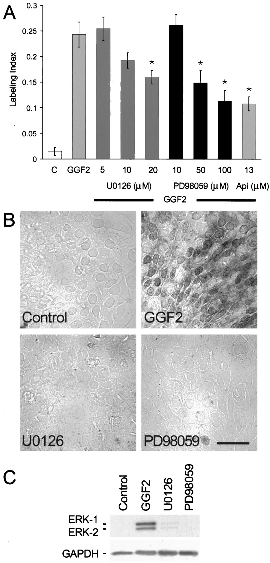
Inhibitors of the ERK–MAPK cascade resulted in moderate reductions in the level of S-phase entry induced by rhGGF2.A, U0126 at 5 and 10 μm did not significantly reduce the level of S-phase entry induced by rhGGF2, but 20 μm reduced the response by 34% (p < 0.05). The MEK1 inhibitor PD98059 at 10, 50, and 100 μm produced a dose-dependent reduction of rhGGF2-induced S-phase entry. The ERK–MAPK inhibitor apigenin (Api) at 13 μm produced approximately the same level of reduction observed for PD98059 at 100 μmwhen tested in the presence of rhGGF2. B, Immunocytochemical analysis with an antibody recognizing activated ERK-1 and ERK-2 showed inhibition of phosphorylated forms of ERK-1 and ERK-2 in the presence of U0126 and PD98059. When pieces of utricular epithelium were treated with 20 μm U0126 or 100 μm PD98059 in the presence of rhGGF2 and then processed for immunocytochemistry, levels of phosphorylated ERK-1 or ERK-2 were virtually undetectable, similar to the control cultures. By comparison, pieces treated for 1 hr with rhGGF2 exhibited strong cytoplasmic and nuclear staining for the phosphorylated forms of ERK-1 and ERK-2. C, Immunoblot analysis of pieces of sensory epithelium that were treated with U0126 or PD98059. Phosphorylated forms of ERK-1 and ERK-2 were readily detected in samples treated with rhGGF2 (lane 2) but were nearly undetectable in samples treated with rhGGF2 in the presence of 20 μm U0126 (lane 3) or 100 μm PD98059 (lane 4) or in control cultures (lane 1). Scale bar, 50 μm.


