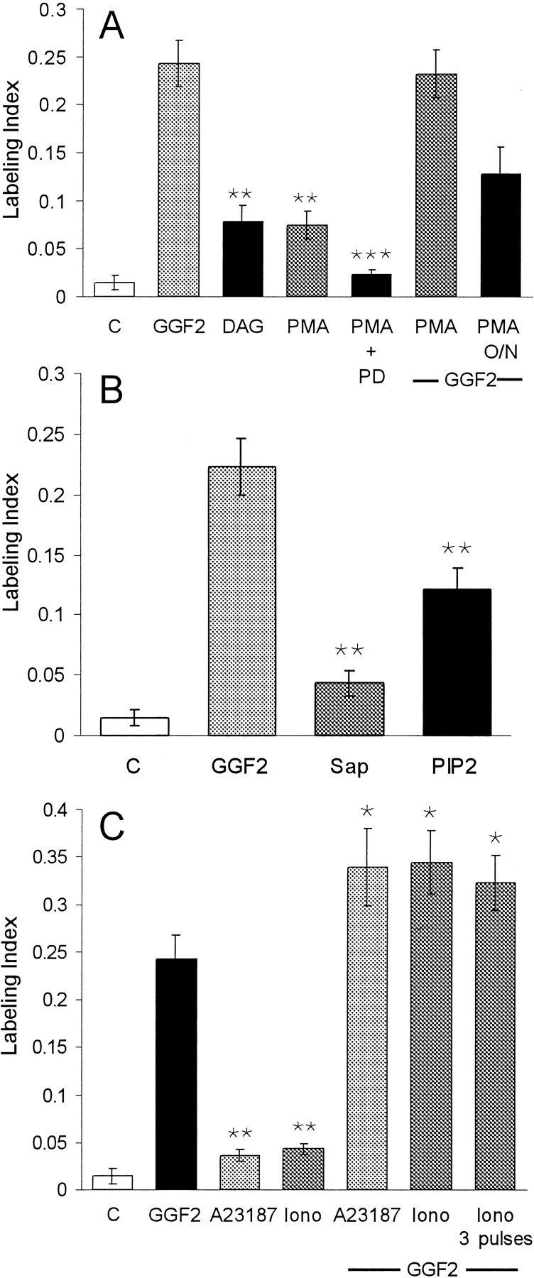
Brief treatments with activators for PKCs, PKB–Akt, and increased intracellular calcium induced S-phase entry in utricular epithelia. A, When pieces of sensory epithelium were treated for 15 min with either 10 μm DAG or 5 pm PMA and then cultured in the standard medium for 72 hr, S-phase entry increased to approximately five times the level seen in control cultures. To inhibit signaling via the ERK–MAPK pathway, other pieces of epithelium were pretreated for 60 min with 50 μm PD98059 (PD) before the 15 min treatment with PMA. The medium was then changed to one containing 50 μm PD98059 and BrdU for the remainder of the 72 hr culture period. Inhibition of the ERK–MAPK pathway significantly reduced the level of S-phase entry induced by the PMA treatment. Other pieces of epithelium were treated with 5 pm PMA for 15 min and subse- quently changed to a medium that contained rhGGF2 for 72 hr. The sequential treatment with PMA and rhGGF2 did not increase the level of S-phase entry over that induced by rhGGF2 alone. To downregulate PKC activity, other cultures were incubated with 5 pm PMA for 16 hr [PMA overnight (O/N)] before rhGGF2 was added for the remaining 56 hr. This resulted in a 32% decrease in the level of labeling compared with that expected for 56 hr in rhGGF2 alone.B, Pieces of sensory epithelium were permeabilized by a 1 min treatment with 10 ng/ml saponin (Sap). Next, half of the pieces were changed to standard medium and cultured for 72 hr. The other half were changed to 2 μm PI-3,4-P2 (PIP2) for 15 min and then changed into standard medium and cultured for 72 hr. Compared with controls, the brief treatment with PI-3,4-P2 resulted in a ninefold increase in S-phase entry measured over the subsequent 72 hr culture period in the presence of BrdU. This was ∼50% of the level that was induced during 72 hr in the continuous presence of rhGGF2. C, When pieces of sensory epithelium were treated with either A23187 or ionomycin (Iono) at 100 nm for just 15 min and then cultured for 72 hr in the standard medium, a level of S-phase entry was induced that was 2.4 and 2.9 times the level that was measured in controls. When other pieces of epithelium were treated with A23187 for 15 min and then cultured for 72 hr with rhGGF2 and BrdU, a level of S-phase entry was observed that was 139% of the level induced by rhGGF2 (142% in the case of ionomycin). Treatments with three 15 min pulses of ionomycin (1 pulse per hour) followed by 72 hr in culture in the presence of rhGGF2 and BrdU did not produce an increase over the level measured for a single 15 min pulse. **Significance compared with control cultures (p < 0.05); ***Significance compared with cultures treated for 15 min with PMA alone (p < 0.05).


