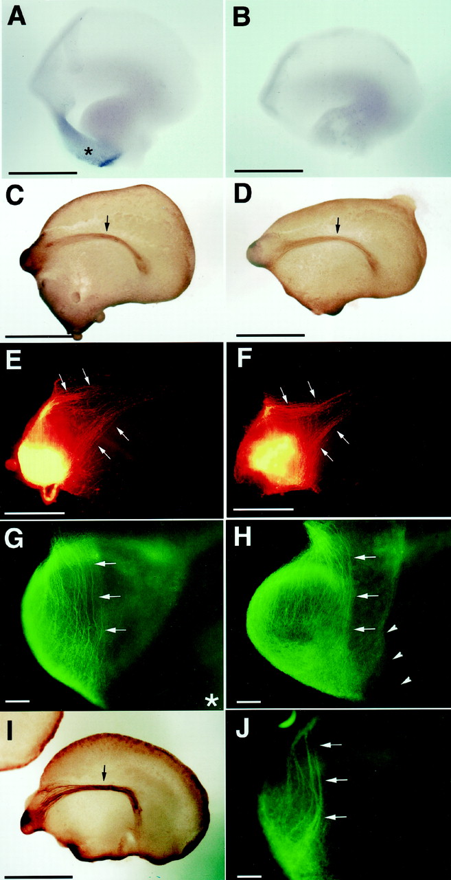
Effects of septum and inhibition of endogenous Slit signaling on mitral cell projection. A,B, Expression of mouse Slit2 mRNA in the E12.5 whole-telencephalon preparation containing the septum (A) and free of the septum (B). Medial views of the preparations. Rostral aspects are to the left. Signals are only detected in the septum (arrowhead) of A. Slit1 mRNA showed a similar expression pattern (data not shown). The septum-retained (C, E, G) and septum-free (D, F,H) telencephalons were organotypically cultured for 2 d, and projection patterns of mitral cell axons were analyzed by various methods. C, D, Lateral views of telencephalons immunostained with anti-neuropilin-1 antibody. Mitral cell axons form the LOT bundles (arrows) in both telencephalons. E,F, DiI was injected on the medial side of the olfactory bulb and filled medial mitral cell axons. Lateral views of the rostral parts of telencephalons. DiI-labeled axons (arrows) turn around in the olfactory bulb from the underside and join the margin of the LOT on the telencephalon surface in both preparations.G, H, Mitral cell axons on the medial side of the olfactory bulb visualized with anti-neuropilin-1 antibody. Mitral cell axons (arrows) projected laterally (toward the underside) in a highly orderly manner, regardless of the presence or absence of the septum. A part of the septum is seen in thebottom right corner in G(asterisk). Arrowheads inH indicate the cut edge from which the septum was removed. RoboN was added to cultures of septum-free preparations (I, J). I, Lateral view of the telencephalon immunostained with anti-neuropilin-1 antibody as in C and D. J, Medial view of the olfactory bulb immunostained with anti-neuropilin-1 antibody as in G and H. In all of the 11 preparations examined, the projections of axons (arrows) are similar to that in the other cultures. Scale bars:A–D, I, 1 mm; E,F, 500 μm; G, H,J, 100 μm.


