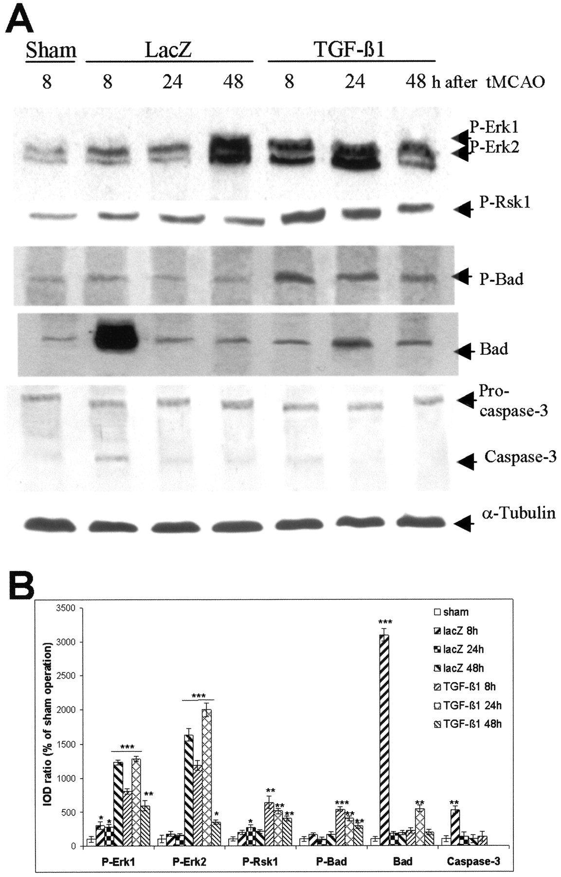
Adenovirus-mediated TGF-β1 overexpression activates Erk1/2 and Rsk1 and interrupts apoptotic cascades through modulation of Bad expression and inhibition of caspase-3 activation in ischemic mice. A, Representative blots of P-Erk1/2, P-Rsk1 (Thr360/Ser364), P-Bad (Ser112), Bad, caspase-3, and α-tubulin. The mice were subjected to intracerebral injection of either AdRSVlacZ or AdRSVahTGF-β1. Thirty minutes of MCAO or sham operation was performed on the fifth day after the injection. The striatal tissues were isolated, and proteins were extracted at the defined time points after ischemia. Sixty micrograms of total protein were loaded to each lane for Western blot. Blots were representative for three animals for each time point. B, Semiquantification of blots. The ratio of IOD from P-Erk1/2, P-Rsk1, P-Bad, Bad, and caspase-3 signals to α-tubulin signals was calculated. The ratios obtained from sham-operated mice were normalized to 100%, and the data were given as means ± SD of the percentage of controls. Caspase-3 activation was not detectable in TGF-β1-transduced mice at 24 and 48 hr after tMCAO. *p < 0.05; **p< 0.01; ***p < 0.001 compared with sham-operated mice.


