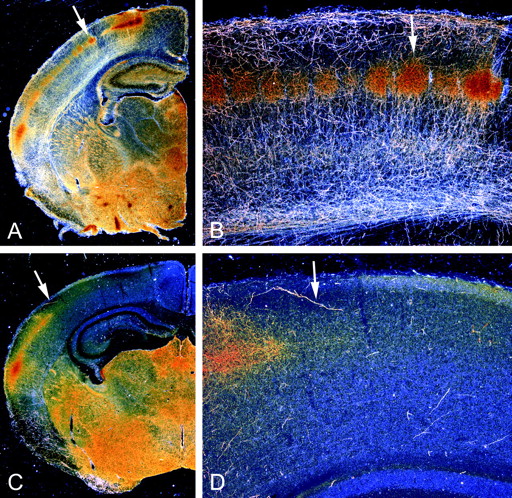
The outgrowth of 5-HT axons to the dorsal neocortex and hippocampus falters in the absence of GAP-43. Dark-field photomicrographs show the distribution of 5-HT axons in coronal sections from P7 mice; arrows demarcate the same regions in low- and high-magnification views. In WT mice (A, B), 5-HT axons are present throughout the brain and densely innervate the thalamus, basal forebrain, hippocampus, and cortex. Distinct patches of serotonin immunoreactivity that demarcate barrels in layer IV of the parietal cortex are visible (arrows in A, B). In GAP43−/− mice (C, D), the growth of 5-HT axons into subcortical regions is relatively normal, but only a few 5-HT axons are observed beyond the ventral aspects of the neocortex.


