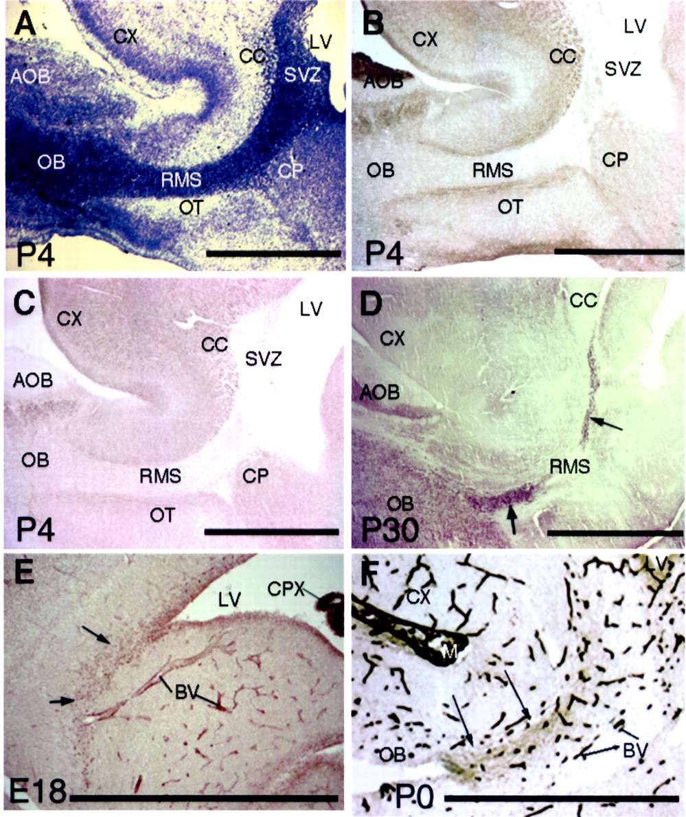
N-CAM, PSA, and laminin expression in the rostral migratory stream. A, Nissl staining of a sagittal section from a postnatal day 4 rat forebrain. The RMS, which is characterized by a high cellular density, begins at the anterior portion of the SVZ and ends at the center of the olfactory bulb (OB). B, C, N-CAM and PSA in the RMS of early postnates. B and C are adjacent sections from a P4 rat forebrain that are stained with anti-N-CAM (clone 5B8) and anti-PSA (clone MenB) MABs, respectively. The SVZ and the RMS stain very weakly, whereas the borders of the RMS show strong staining. D, PSA immunostaining of a P30 mouse forebrain. In contrast to weak expression of PSA in P4 RMS shown inC, PSA is expressed strongly in the RMS (arrows) and the olfactory bulb (OB).E, α5 subunit of laminin in the RMS (arrows) from an embryonic day 18 mouse shows punctate staining. Relatively strong staining is also seen in the choroid plexus (CPX) of the lateral ventricle (LV) and blood vessels (BV). F, γ1-Laminin is expressed in the RMS (arrows) from a P0 rat brain. Blood vessels (BV) and the meninges (M) are also stained. Scale bars:A–D, F, 1 mm; E, 50 μm.AOB, Accessory olfactory bulb; CC, corpus callosum; CP, caudate-putamen; CX, cerebral cortex; OT, olfactory tubercle.


