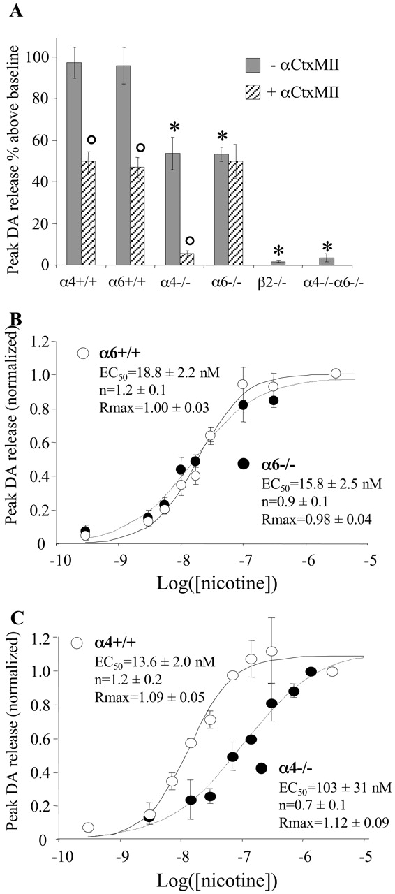
Nicotine-induced DA release in striatal synaptosomes. A, Striatal synaptosomes from α4-/-, α6-/-, β2-/-, and α4-/-α6-/- animals and their respective Wt controls were prepared and loaded with 3H-DA. The effect of 3 μm l-nicotine on DA release was determined in the presence or absence of 100 nm αCtxMII. Peak 3H-DA release values (expressed as percentage of release above baseline levels) represent the mean ± SEM of at least three determinations. *p < 0.01 compared with matching Wt control (Student's t test); °p < 0.01 compared with αCtxMII-free condition (paired Student's t test). B, C, Dose-response curves for nicotine-induced DA release. For each synaptosome preparation, data were normalized to the response obtained with 3 μm nicotine. Dose-response curves were fitted to the Hill equation: Release = Rmax/(1+(EC50/[Nic])n).


