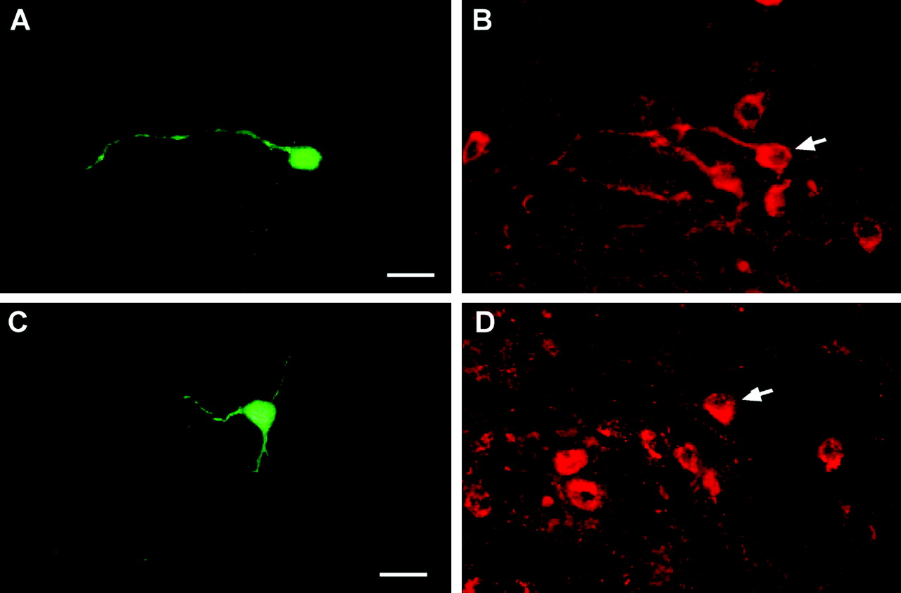
Figure 2.
Double labeling of dopamine and POMC neurons. Representative dopamine (A, B) and POMC (C, D) neurons that responded to estrogen. Arcuate neurons were filled with biocytin during the whole-cell recording. A, Biocytin-streptavidin-Cy2 labeling of fusiform arcuate neuron. Scale bar, 20 μm. B, Immunocytochemical staining of TH in the same neuron (arrow). C, Biocytin-streptavidin-Cy2 labeling of a small pyramidal arcuate neuron. Scale bar, 20 μm. D, Immunocytochemical staining of β-endorphin in the same neuron (arrow).


