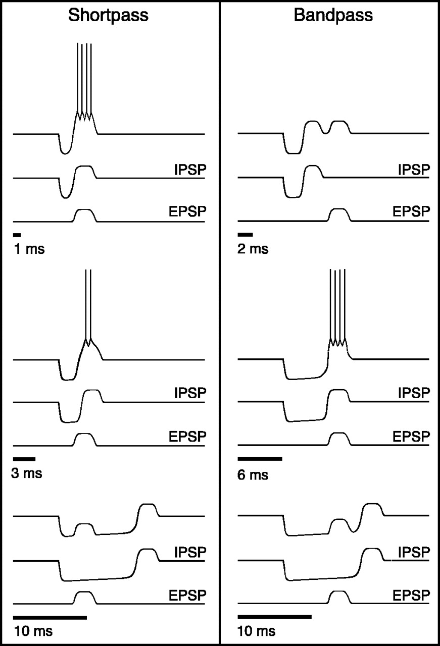
Model for the creation of short-pass and bandpass duration tuning. Above each stimulus (black bars), threetraces are shown. The middle andbottom traces represent hypothetical synaptic inputs to an IC neuron; the top traces show the resulting change in the membrane potential of the cell and, if suprathreshold, its spike output. The model has three components: (1) transient, onset-evoked, subthreshold EPSP (bottom traces); (2) sustained, onset-evoked, IPSP; and (3) a following transient, excitatory rebound from inhibition (middle traces). Neurons fire only when the EPSP coincides with the excitatory rebound from the IPSP. The best duration and range of duration selectivity of a cell are determined by the duration of the EPSP and its latency relative to the IPSP.Shortpass, For the 1 msec tone, the EPSP coincides with the rebound from inhibition, and the summation pushes the membrane potential of the cell above the spike threshold. For the 3 msec tone, the EPSP is partially cancelled by the IPSP, resulting in a weaker suprathreshold response and a longer first spike latency. For the 10 msec tone, the EPSP occurs before the end of the IPSP and is rendered subthreshold by the sustained inhibition. Bandpass, For the 2 msec tone, the rebound from inhibition occurs before the EPSP. For the 6 msec tone, the rebound from inhibition coincides with the EPSP, and the summation pushes the membrane potential of the cell above spike threshold. For the 10 msec tone, the EPSP occurs before the end of the IPSP and is rendered subthreshold by the sustained inhibition. Modified from Casseday et al. (2002).


