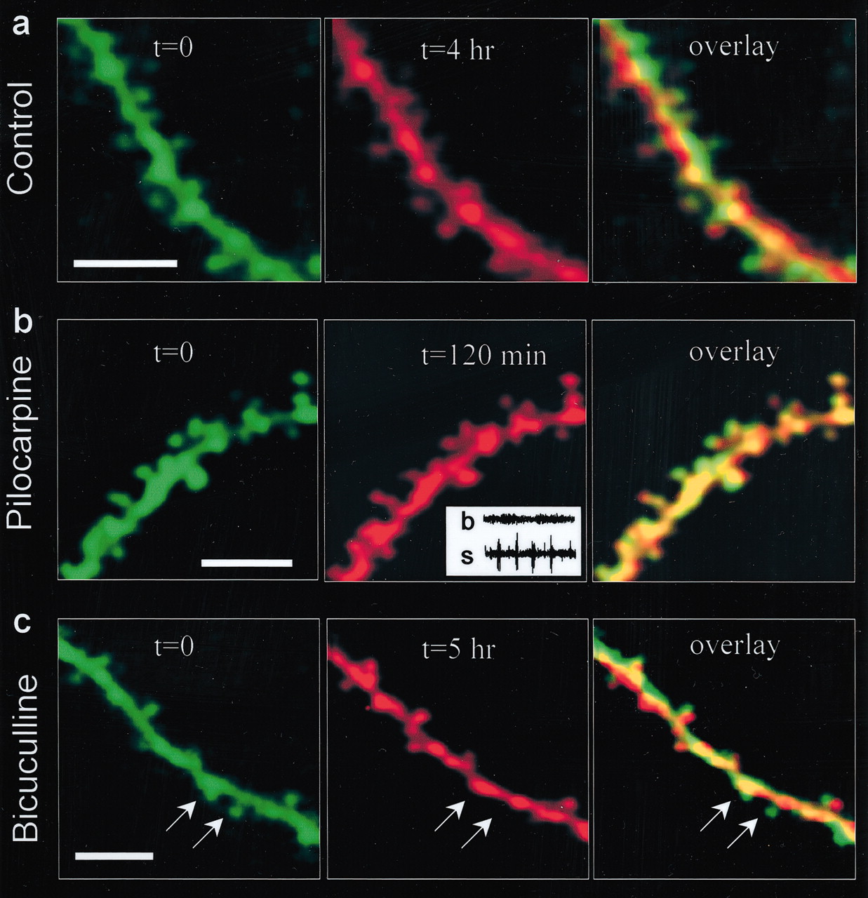
Time-lapse imaging of spines in CA 1 hippocampal neurons in vivo. a–c, Representative examples of spiny dendrites from CA 1 basal dendrites under control conditions (a), during pilocarpine-induced seizures (b), and after application of bicuculline (c). The first imaging session (t = 0, pseudocolored green) is compared with the second imaging session (pseudocolored red) by overlying the two images (overlay). The overlays indicate little change in spines. However, rare instances of spine loss were observed after bicuculline treatment (arrows in c). Inset in b, 10 sec traces from the EEG recordings before (b) and during the seizure (s). All images are single optical slices. Scale bars, 5 μm.


