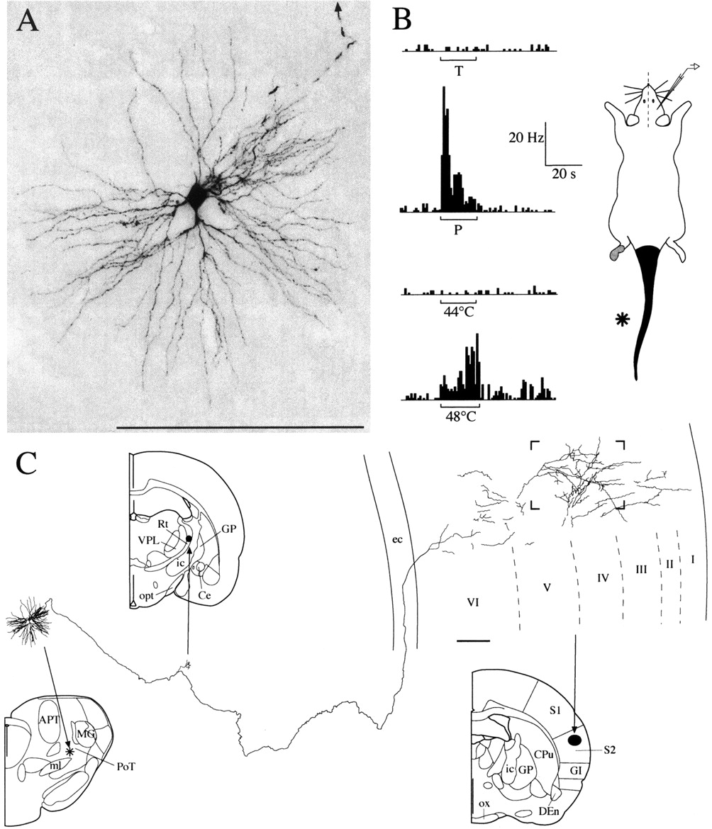
Nociceptive specific PoT neuron j42 labeled with juxtacellular injection of biotin-dextran. A, Microphotograph of the labeled soma with its dendrites and first portion of the axon (arrow at the top). B, Top to bottom, Response to innocuous touch (T), noxious pinch (P), innocuous thermal (44°C), and noxious thermal (48°C) stimuli, applied for 20 sec (bars) to the preferential receptive field (area in black plus asterisk); in gray, receptive field area inducing moderate firing. C, Reconstruction of the neuron from serial coronal sections. Left to right, Coronal levels are 4.20, 6.44, and 7.70 mm interaural (Paxinos and Watson, 1998). Note: from left, (1) the soma plus dendrites in PoT, (2) a slight axonal arborization in the reticular thalamic nucleus (Rt), and (3) the extensive axonal arborization in S2 cortex. The framed region in cortex is illustrated in the high-magnification photomicrograph in Figure 8. Abbreviations are listed in Figure 1 legend. Scale bars, 200 μm.


