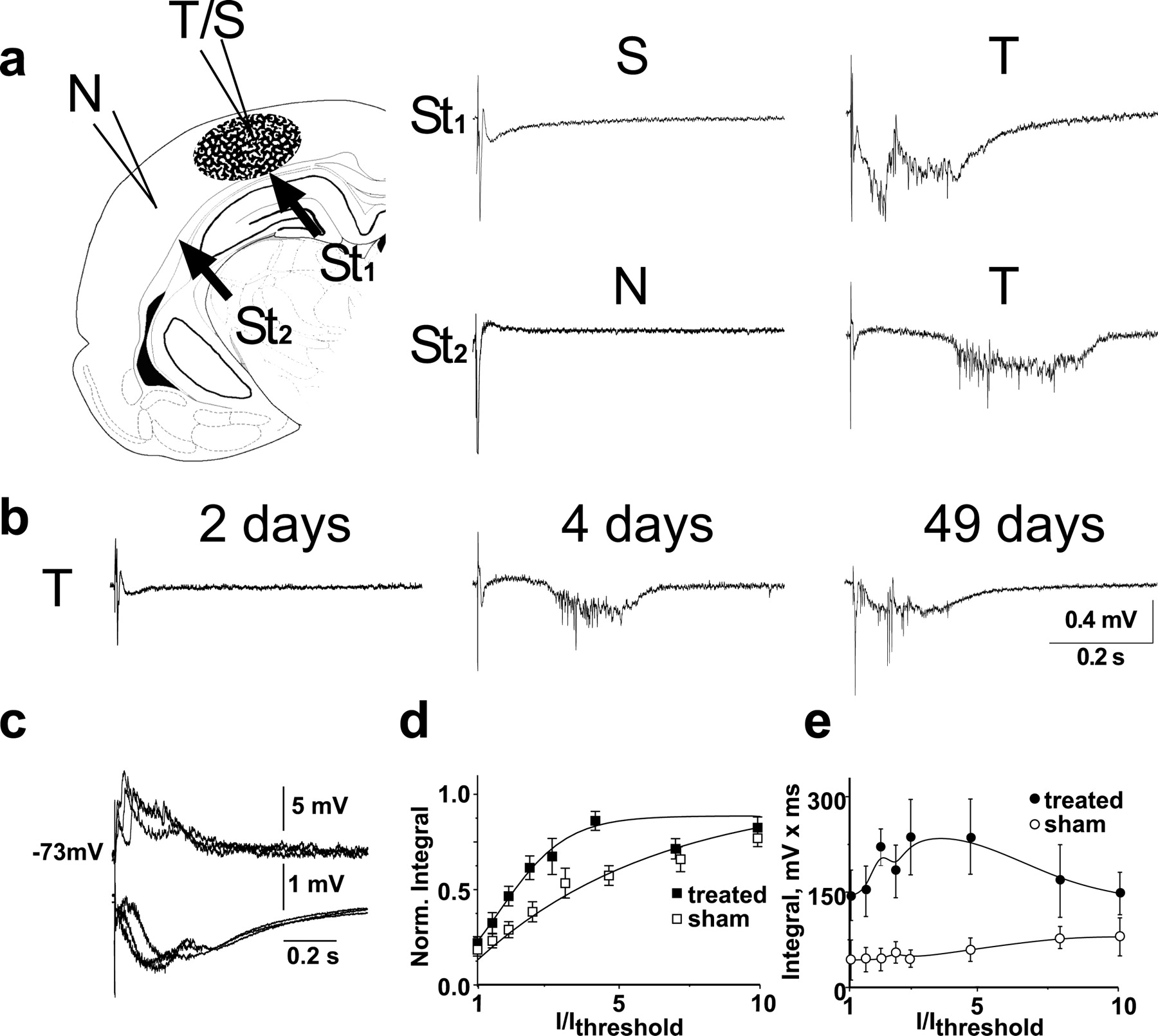
Electrophysiological responses in BBB-disrupted cortex. a, Electrophysiological recordings were performed in slices from BBB-treated as well as nontreated cortical regions from operated and sham-operated animals. In the BBB-treated region the typical potential evoked in response to white matter stimulation (St1) showed an early, short-duration potential followed by a late, long-duration, event with a variable latency. Stimulation in the white matter outside the treated region (St2) often elicited delayed paroxysmal activity limited to the treated region. b, Paroxysmal events were first recorded in slices 4 d after treatment. Scaling refers to a and b. c, Overlapping traces in response to three consecutive extracellular stimuli (0.1 Hz); extracellular (bottom) and intracellular recordings (top) show that synaptic paroxysmal responses are evoked synchronously. Interelectrode distance was ∼300 μm. d, e, The integral of the early (d, <50 msec after stimulus artifact, values for each slice were normalized to the maximal response) and late (e, 50-500 msec) evoked responses in slices from treated (filled symbols) and sham-operated (open symbols) cortices under increasing stimulus intensity (normalized to threshold intensity: Ithreshold). T, Treated; S, sham-operated; N, nonoperated.


