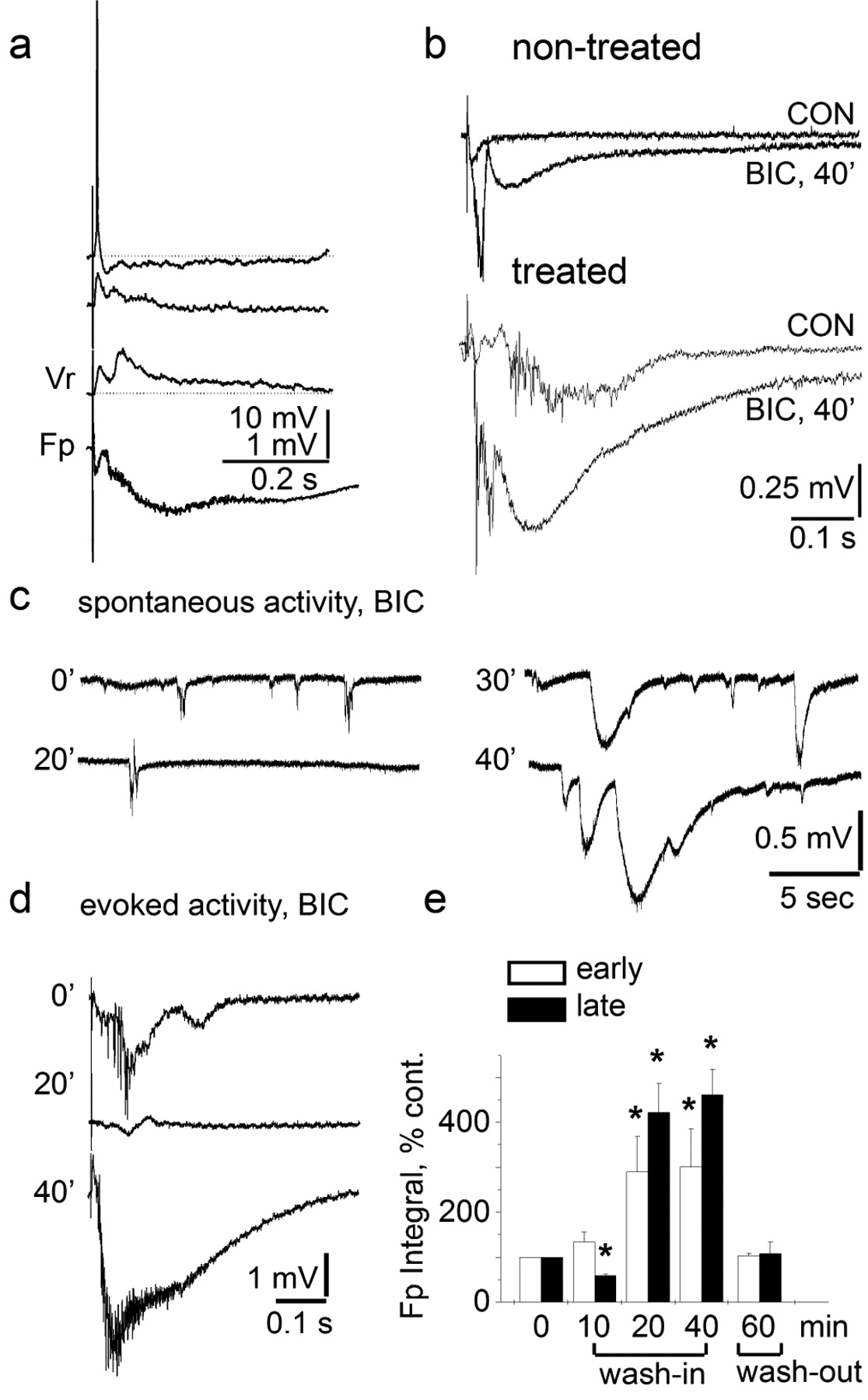
Paroxysmal activity is associated with GABA-mediated inhibition: a, Simultaneous intracellular (top traces) and extracellular recordings (Fp, bottom traces) are shown in response to extracellular stimulation. Responses to consecutive stimulation are shown at different membrane voltages in relation to the resting potential (Vr = -70 mV). b, Simultaneous extracellular recordings from the nontreated and treated somatosensory cortex of the same slice. Superimposed traces are shown under aCSF (CON) and 40 min after the addition of bicuculline (BIC). Note the enhanced negative deflection in the field potential recorded in the treated region of the slice. c, Recordings from a slice with spontaneous interictal-like epileptiform activity during perfusion of bicuculline-containing aCSF. Note the initial suppression followed by increase in frequency of appearance and amplitude of spontaneous activity. d, Extracellular recordings during bicuculline perfusion were associated with an initial reduction in duration and amplitude of paroxysmal activity (20′) followed by the appearance of large field potential (40′). e, Quantitative analyses of the integral of early and late evoked responses under bicuculline (n = 4; see Results). *p < 0.05 (in comparison to control values).


