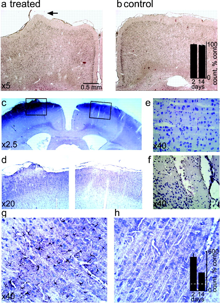
Focal cortical perfusion with DHC or albumin caused astrocytic activation with no neuronal loss. a, b, Whereas histological sections 24 hr after treatment show slight extracellular edema in the treated hemisphere, cortical structure is otherwise normal. Arrow points to the treated, exposed cortical region. Bar graphs represent cell count from histological sections 2 and 14 d (n = 6 each) after treatment in percentages compared with the contralateral nontreated region. c, d, Coronal sections 48 hr after perfusion with albumin, showing normal cortical structure under low magnification. The black boxes represent the cortical regions seen under a larger magnification in d. e, In a large magnification normal cortical structure is observed. Note a typical cortical capillary showing blood cells limited to the vessellumen. f, For comparison, 24 hr after a traumatic cortical injury extravasation of blood cells is observed in the cortical tissue (for methodological details, see Eyupoglu et al., 2003). g, h, A prominent increase in the number of GFAP-stained astrocytes is seen in an albumin-treated cortex compared with the nontreated contralateral cortex. Bar graphs represent counted GFAP-positive cells (in percentage to the contralateral hemisphere) from histological sections 2 and 14 d (n = 6 each) after treatment.


