Nitric Oxide Synthase (NOS)-Interacting Protein Interacts with Neuronal NOS and Regulates Its Distribution and Activity
Supplemental data
Files in this Data Supplement:
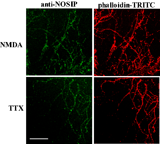
- supplemental material - Supplementary fig. 1: Anti-NOSIP immunoreactivity in terminal dendrites of cultured neurons acutely treated with NMDA (1 �M) and glycine (10 �M) or tetrodotoxin (TTX, 0.5 �M). Phalloidin-TRITC labelling was used to counterstain the actin cytoskeleton. Scale bar represents 40 �m.
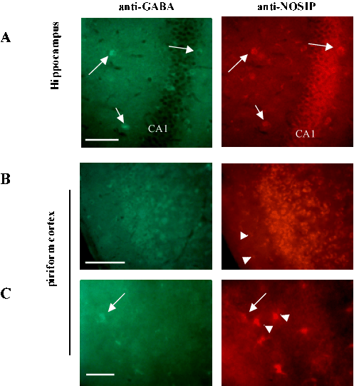
- supplemental material - Supplementary fig. 2: Colabelling of brain sections of mice treated with 25 mg/kg kainate with anti-NOSIP and anti-GABA antibodies. (A) Staining of interneurons in the hippocampal molecular layer (arrow points to a co-labelled cell). (B and C) Staining of piriform cortex with anti-GABA antibody reveals co-labelling of interneurons (arrow) with anti-NOSIP antibody, but not of additional, NOSIP-positive cells abundant in layer I, which resembled stellate astrocytes in morphology (arrowheads).� CA2 � CA2 sector of the pyramidal cell layer; I � cortical layer I. Scale bars represent 200 �m in A and B and 40 �m in C.
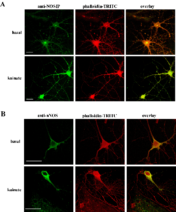
- supplemental material - Supplementary fig. 3: Changes in immunoreactivity for NOSIP (A) and nNOS (B) in cultured neurons at 40 h following treatment with kainate (20 �M). Phalloidin-TRITC was used to counterstain the actin cytoskeleton. Scale bars represent 20 �m in A and B.

- Print ISSN: 0270-6474
- Online ISSN: 1529-2401
