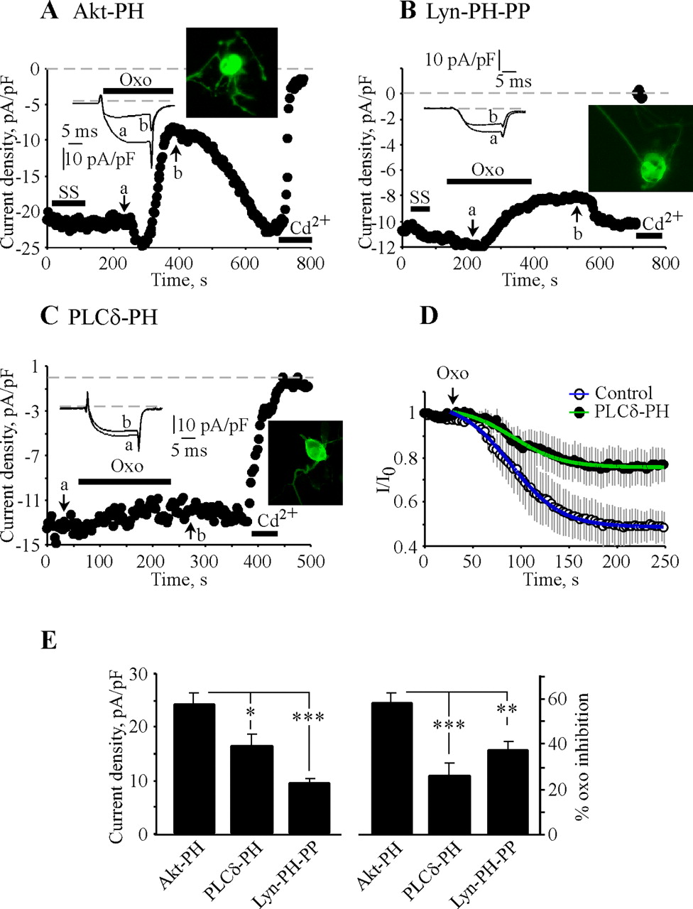
Overexpression of PIP2 depleting-sequestering constructs in SCG neurons decreases ICa density and reduces muscarinic suppression. A-C, PTX-treated SCG neurons were studied in perforated-patch configuration. Voltage protocols and other experimental conditions and labeling are as in Figure 2. Cells were transfected using the biolistic particle delivery system (gene gun; see Materials and Methods for details) with Akt-PH (A), Lyn-PH-PP (B), or PLCδ-PH (C). Color insets on the right show images of the transfected neurons. Cd2+, CdCl2. D, Pooled time courses of muscarinic suppression of ICa in Akt-PH-(white circles; n = 8) and PLCδ-PH-transfected (black circles; n = 8) cells, temporally aligned to the time of oxo application. Data were fitted by the equation y = y0 + a/(1 + exp(-(t1/2 - t)/k)), where y is I/Io, a is ymax - ymin, t1/2 is a half-time of inhibition, and k is a slope factor. For Akt-PH-expressing cells, y0, a, t1/2, and k were 0.01, 1.1, 60.4, and -26.3, respectively; for PLCδ-PH-expressing cells, they were 0.02, 1.1, 62.2, and -25.6, respectively. E, Bars on the left summarize data of the current density of the Akt-PH-, Lyn-PH-PP-, and PLCδ-PH-expressing neurons (n = 18, 23, and 18, respectively), and those on the right summarize their muscarinic modulation (n = 10, 12, and 10, respectively). Significance: *p ≤ 0.05; **p ≤ 0.01; ***p ≤ 0.001.


