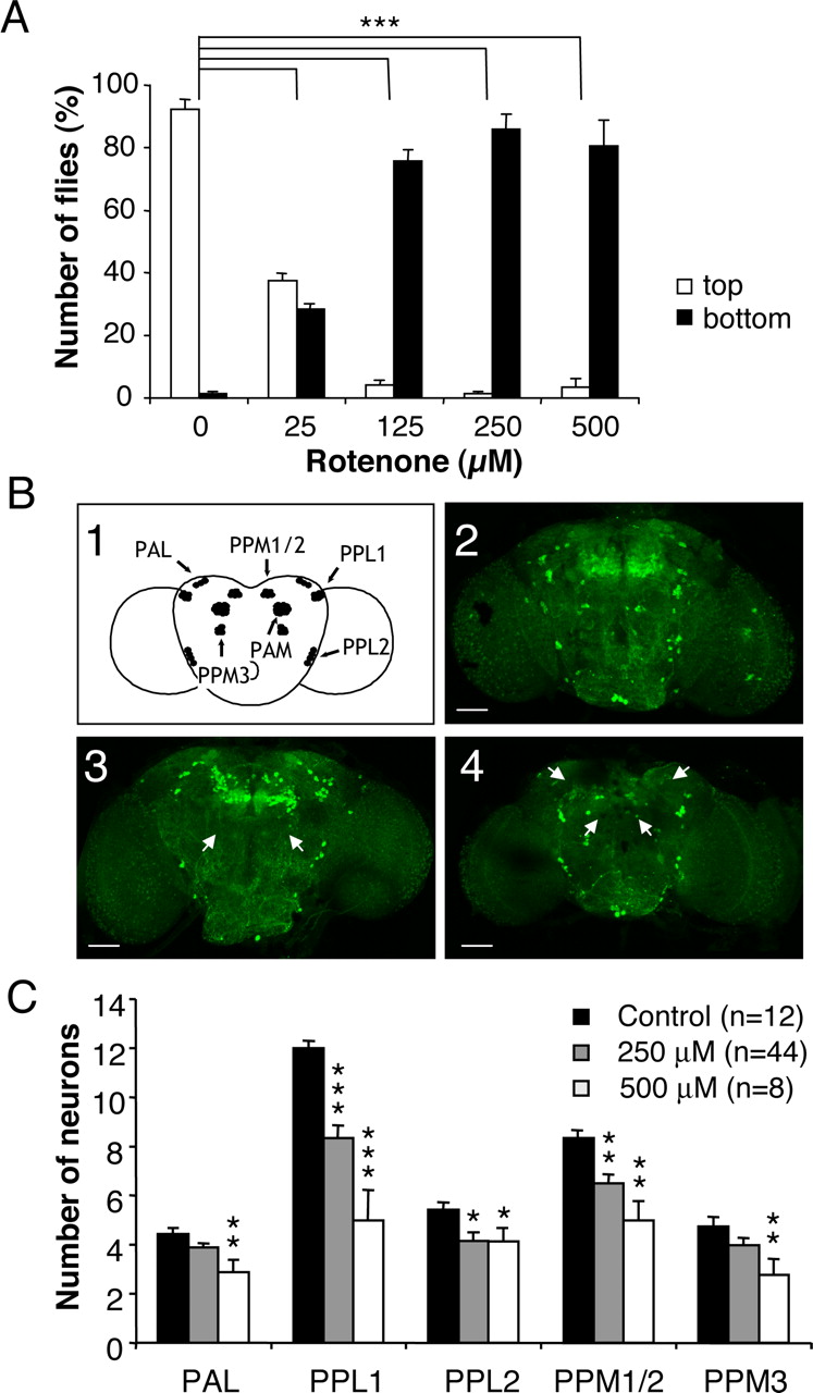
Exposure of Drosophila to rotenone induces severe locomotor deficits and dopaminergic neuron loss. A, Negative geotaxis assay of adult flies exposed previously for 7 d to various amounts of rotenone. White bars indicate the percentage of flies that climbed to the top of the column, and black bars indicate the percentage of flies that remained at the bottom after 1 min. Differences in PI between control and rotenone-treated flies were highly significant (***p < 0.001). B1, Schematic representation of the dopaminergic neuron clusters in Drosophila adult brain in frontal view. B2-B4, Tyrosine hydroxylase immunolabeling showing dopaminergic neuron patterns in multifocal confocal views of adult fly brains after 7 d of exposure to 0 (control), 250, and 500 μm rotenone, respectively. White arrows indicate the total absence of certain clusters after rotenone exposure (PPM3 in B3 and PAM and PAL in B4). Scale bars, 50 μm. C, Quantification of the number of neurons in dopaminergic clusters of control brains (black bars) or in brains of flies exposed for 7 d to 250 μm (gray bars) or 500 μm (white bars) rotenone. The density of the neurons in the PAM clusters was too high to allow precise scoring of their number. n indicates the number of brain hemispheres examined in each condition. *p < 0.05, **p < 0.01, and ***p < 0.001 compared with control values.


