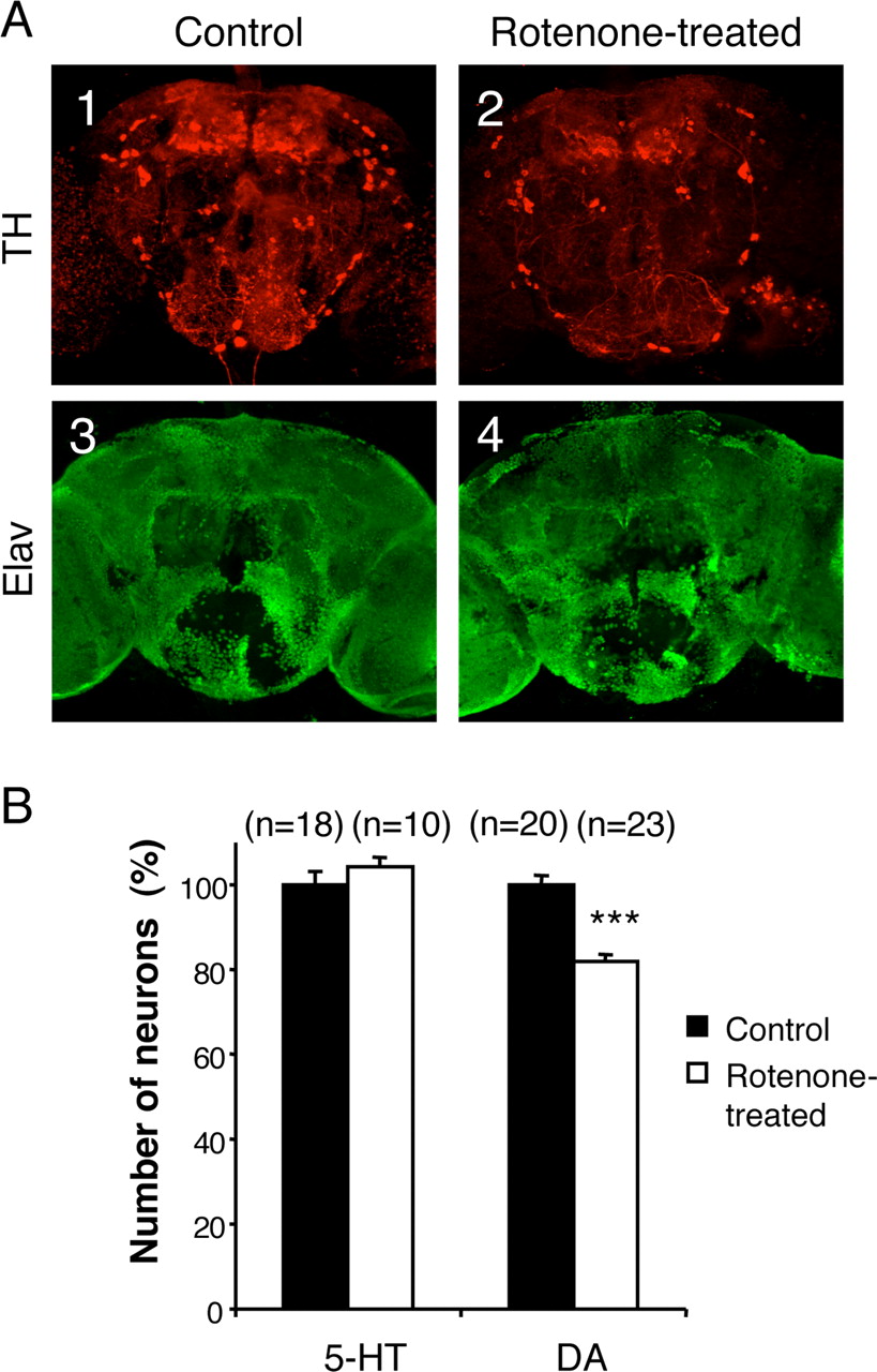
Figure 2.
Selectivity of the neuronal degeneration induced by rotenone. A, Coimmunolabeling of adult brains with anti-tyrosine hydroxylase (TH) (A1, A2) and anti-Elav (A3, A4) antibodies after 7 d of exposure to 125 μm rotenone. The pesticide induced obvious dopaminergic neuron loss, whereas the general pattern of the pan-neuronal Elav marker was not modified. B, Total number of serotoninergic (5-HT) and dopaminergic (DA) neurons in fly brains exposed for 7 d to 125 μm rotenone (white bars) compared with control brains (black bars), expressed as a percentage of the mean value of the control. n indicates the number of brain hemispheres examined in each condition. ***p < 0.001.


