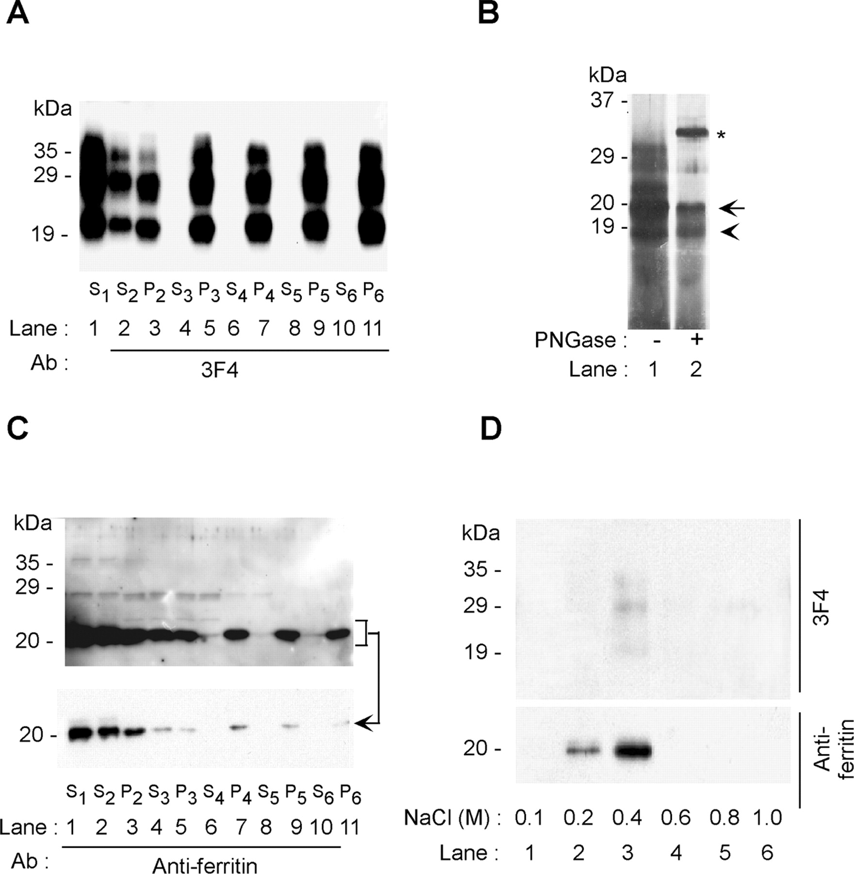
Protease-resistant sCJD-PrPSc is associated with ferritin. Clarified supernatant obtained from PK-treated CJDH (S1) was subjected to five additional rounds of ultracentrifugation, and the resulting detergent-soluble supernatant (S2-S6) and detergent-insoluble pellet (P2-P6) fractions were immunoblotted with 3F4. A, The starting material (S1) and the supernatant and pellet fractions obtained after ultracentrifugation (S2 and P2, respectively) show the N-terminally truncated PrPSc glycoforms migrating at 29 and 30, 22-25, and 19 kDa, respectively (lanes 1-3). After the first round of ultracentrifugation, 60% of PrPSc fractionates in the pellet fraction (lanes 2, 3). After subsequent rounds of ultracentrifugation, all of the PrPSc is detected in the pellet fractions (lanes 4-11). B, Silver staining of the P6 fraction shows PrPSc glycoforms of 19-30 kDa and a 20 kDa band (lane 1). After treatment with PNGase, all PrPSc glycoforms migrate at 19 kDa, as expected (lane 2, arrowhead), whereas the 20 kDa band remains insensitive to deglycosylation (lane 2, arrow). The slower migrating band in lane 2 (*) is derived from the added PNGase. C, Reprobing of the membrane in A with anti-ferritin antibody shows strong immunoreaction with the 20 kDa band and minor species migrating at 25 and 30 kDa (lanes 1-11). The 20 kDa band is detected in the first three fractions (lanes 1-3) and in all subsequent pellet fractions (lanes 4-11). D, The P6 fraction from A was treated with NaCl and subjected to DEAE-cellulose chromatography. Immunoblotting of eluted proteins with 3F4 and anti-ferritin antibodies shows dissociation of ferritin from PrPSc at concentrations of 0.2-0.4 m NaCl (lanes 2, 3).


