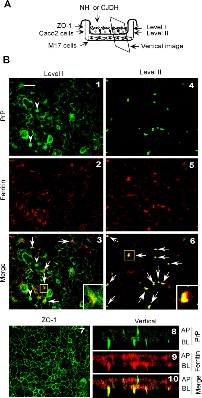
sCJD-PrPSc is cotransported with ferritin across Caco-2 cells. A, Diagrammatic representation of filter inserts with Caco-2 cell monolayers and the levels at which images were captured. M17 cells were cultured on glass coverslips in the BL chamber to capture transcytosed PrPSc-ferritin complexes (see Fig. 7A). B, Caco-2 cell monolayers were incubated with CJDH-DE in the AP chamber for 2 hr, and the filters were cut into two pieces. One was immunostained with 8H4-anti-mouse FITC (green) and anti-ferritin-anti-rabbit RITC (red) (panels 1-6), and the other was immunostained with with anti-ZO-1-anti-rabbit FITC (green) (panel 7). A horizontal image at level I shows colocalization of PrP (green) and ferritin (red) on the AP surface of Caco-2 cells (panels 1-3, arrows). Some PrP immunoreaction without ferritin is also seen (green) (panels 1-3, arrowheads). A horizontal image captured at level II shows colocalization of PrP (green) and ferritin (red) at the filter pores (panels 4-6, arrows). Immunostaining for ZO-1 shows the presence of tight junctions throughout the monolayer (panel 7). A vertical section through the Caco-2 monolayer shows similar colocalization of PrP (green) and ferritin (red) at the AP and BL membranes, as observed in the horizontal images (panels 8-10). Scale bar: 10 μm; inset, 2.5×.


