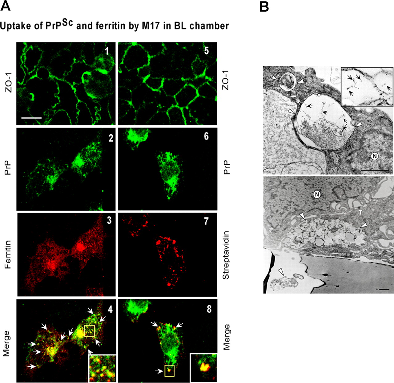
sCJD-PrPSc remains associated with ferritin after transcytosis. A, Caco-2 cell monolayers were placed in a 12-well culture dish containing M17 neuroblastoma cells cultured on coverslips in the BL chamber, and biotinylated CJDH-DE was added to the AP chamber (see Fig. 6 A). After an overnight incubation, Caco-2 cells on filters and M17 cells on coverslips were processed for immunostaining. Immunoreaction of filters with anti-ZO-1 shows the presence of tight junctions in all monolayers (green; panels 1, 5). Immunostaining of M17 cells in the BL chamber for PrP (green) and ferritin (red) shows colocalization of the two proteins, indicating the presence of intact PrPSc-ferritin complexes after transcytosis (panels 2-4, arrows). Coimmunostaining of M17 cells for PrP (green) and streptavidin (red) (panels 6-8) confirms that the PrP signal is derived from biotinylated CJDH-DE in the AP chamber. Scale bar, 10 μm. B, Electron microscopic analysis of the PrPSc-ferritin complex immunoprecipitated with 8H4 shows fibrillar structures and membranes decorated with ferritin aggregates (top inset; arrows). When added to Caco-2 cells, the complex is internalized in relatively large phagosome-like structures surrounded by a single membrane (top; arrowheads). Some of these vesicular structures are extruded out from the BL surface of Caco-2 cells and are seen within the pore of the membrane filter (bottom; arrowhead). (The internalized material in phagosomes shows similar structures as observed in the immunoprecipitated material.) Scale bar: 0.25 μm; inset, 1.5×. N, Nucleus; T, tight junction.


