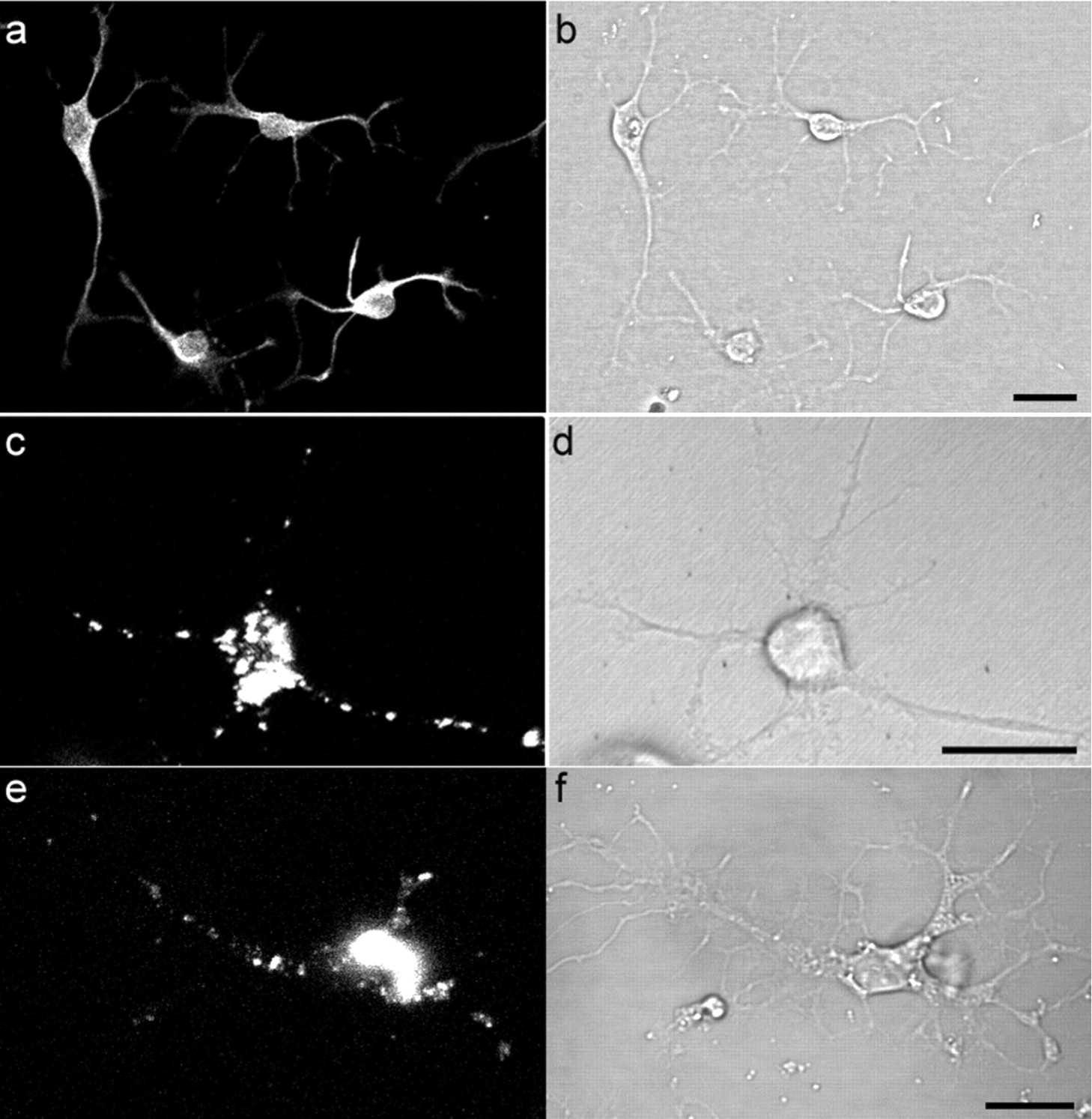
Internalization and trafficking of PrP-resA568 in primary cultured neurons. Cultures derived from the adult hamster brain contained a majority of neuronal cells that were stained with anti-MAP2 (a) with its corresponding differential interference contrast image in b. After 4 d of culture, hPrP-resA568 was added to the hamster cells and imaged at 2 d after exposure to hPrP-resA568. A representative neuron that internalized hPrP-resA568 is shown in c and d. e, m(Ch)PrP-resA568 uptake and trafficking after 2 d of exposure to a neuron cultured from knock-out mice lacking PrP gene expression, with its corresponding differential interference contrast image in f. Scale bars, 20 μm.


