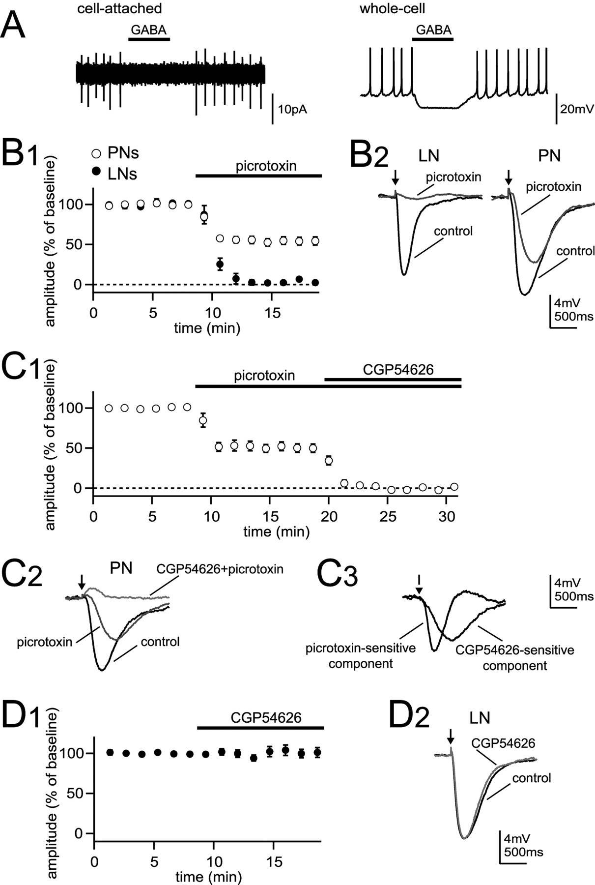
GABA hyperpolarizes antennal lobe neurons via both GABAA and GABAB receptors. A, A 1 s microiontophoretic pulse of GABA at the soma suppresses spontaneous spiking of an LN recorded in cell-attached mode (left). GABA has a similar effect after rupturing the patch (right). B1, GABA was microiontophoresed periodically into the neuropil, and the amplitude of the GABA-evoked hyperpolarization was monitored over time in whole-cell recordings. The graph plots mean ± SEM hyperpolarization amplitude as a percentage of control. Picrotoxin blocked the GABA response in LNs but suppressed less than one-half of the GABA response in PNs. B2, Representative traces show GABA response before and after addition of picrotoxin in an LN (left) and a PN (right). The arrows mark GABA pulses. C1, CGP54626 blocks the picrotoxin-resistant GABA response in PNs. C2, Representative traces show GABA response in a PN before and after addition of picrotoxin and after addition of CGP54626. C3, Subtracted traces show the picrotoxin-sensitive component and CGP54626-sensitive component from traces in C2. D1, CGP54626 does not affect the GABA response in LNs. D2, Representative traces show the GABA response in an LN before and after addition of CGP54626. Error bars represent SEM.


