Cortical Control of Zona Incerta
Supplemental Data
Files in this Data Supplement:
- supplemental material - Supplementary material 1. Firing frequencies of the different ZI cell types recorded under urethane anesthesia, during different cortical states. Unclassified means that the neurons did not have sufficient number of spikes during slow oscillation for categorization or the cortical states did enter slow oscillation during the recordings. �N� refers to the total number of cells in each class (first column) and the number of cells recorded under different cortical states (fast or slow oscillations dominated states or during HVSs) within each class. �Range� refers to the average firing rate of the slowest and fastest neuron during each state. In case of 3 ZI cells (not shown here) only the anatomical data were used.
- supplemental material - Supplementary material 4. Firing frequencies of the different ZI cell types recorded under neuroleptanalgesia, during different cortical states. �N� refers to the total number of cells in each class (first column) and the number of cells recorded under different cortical states (non 5-9 Hz and 5-9 Hz epochs) within each class. �Range� refers to the average firing rate of the slowest and fastest neuron during each state.
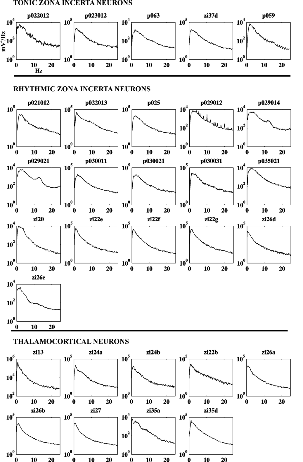
- supplemental material - Supplementary material 2. Power spectra of the LFP in a representative sample of five tonic ZI cells, 16 rhythmic ZI cells and 9 thalamocortical cells during slow cortical oscillations. In all cases lower frequencies (1-3) dominate the spectra.
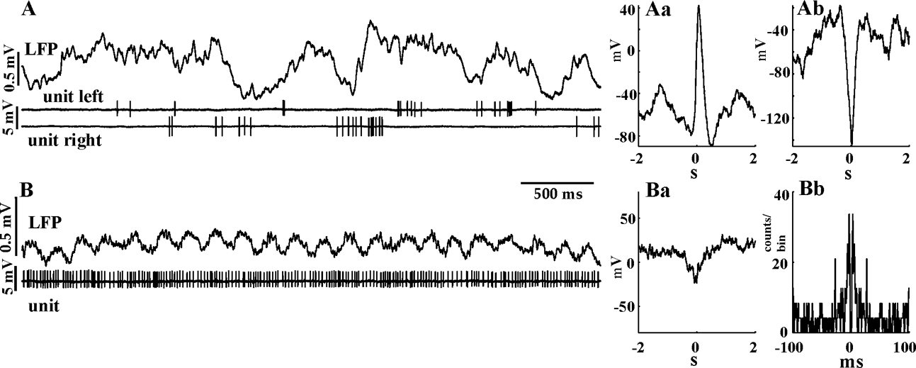
- supplemental material - Supplementary material 3. A) Dual juxtacellular recording of two ZI neurons located in different hemispheres under neuroleptanalgesia. Spike triggered LFP averages of the left (Aa unit l) and right unit (Ab unit r). The activity of both cells is significantly modulated by the slow waves but note the opposite phase relationship of the two cells to the cortical LFP B) A different ZI cell fires in a tonic manner during a low voltage cortical activity STA (Ba) demonstrate weak modulation, whereas the autocorrelogram (Bb), indicate a relatively regular firing.
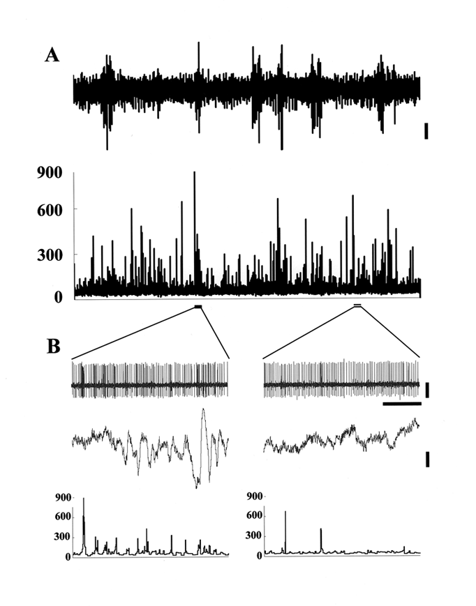
- supplemental material - Supplementary material 5. High frequency spike transients (HFT) embedded in a tonic spike train. A) Upper trace, high pass-filtered EEG signal (1 Hz cutoff frequency), lower part, the instantaneous frequency (IF) function of the unit is shown. Amplitude elevations of the EEG recording mark 5-9 Hz episodes. On the IF plot HFTs can be observed as sudden and robust increase of IF. B) A 5-9 Hz episode (left) and a non 5-9Hz episode (right) are shown at faster sweep (2 sec). Note the higher number of HFTs during 5-9 Hz episodes compared to non-5-9 segments. On the left the weak coupling of HFTs to the co-occurring 5-9 Hz waves can be observed. However, the highest frequency transient precedes the appearance of the 5-9 Hz epoch. On the right two transients detected during non-5-9 Hz EEG activity. Horizontal bar on panel B = 0.5 s, vertical bar on panel A = 100 mV, on panel B for EEG = 100 mV, for unit = 1.5 mV.
- supplemental material - Supplementary material 6. Full, 360� rotation of the 3D reconstructed dendritic trees of a dZI (above; red/yellow) and a vZI (below, blue) neurons around the dorso-ventral axis. The two cells were recorded and labeled in the same track The movie starts with the coronal image from the posterior view, lateral is to the left. The 90� rotation (lateral view, ~5 sec in the movie) and the 270� rotation (medial view, ~15 sec in the movie) reveal the large difference between the mediolateral and anteroposterior extent of both the dZI and the vZI cells.

- Print ISSN: 0270-6474
- Online ISSN: 1529-2401
