Figure 1.
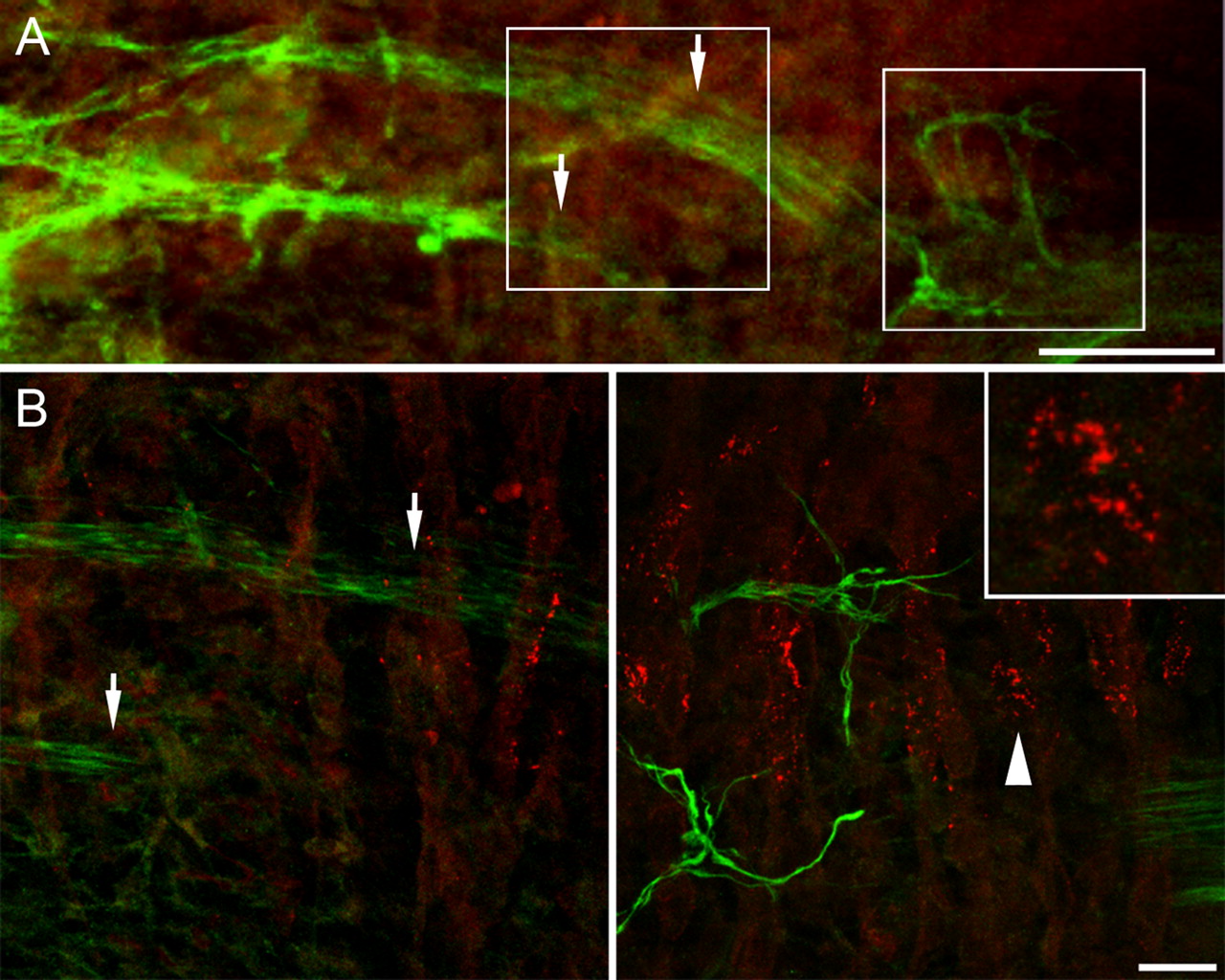
Wild-type mouse diaphragm at E12.5. Maximum intensity projection of stacks of images taken at 2 μm (A) and 0.1 μm (B) intervals. Speckles of AChRs are present on the myotubes near but not distant from the area of nerve entry. They run in a band centered beneath the nerve, but none are in contact with it. Left and right frames in A denote the areas enlarged in left and right bottom B, respectively. Arrows mark nerve trunks in A and B, left. Close-up inset in B, right, shows microclusters grouped but not fused to a single AChR cluster (arrowhead). Scale bars: A, 70 μm; B and inset, 20 and 7 μm, respectively.


