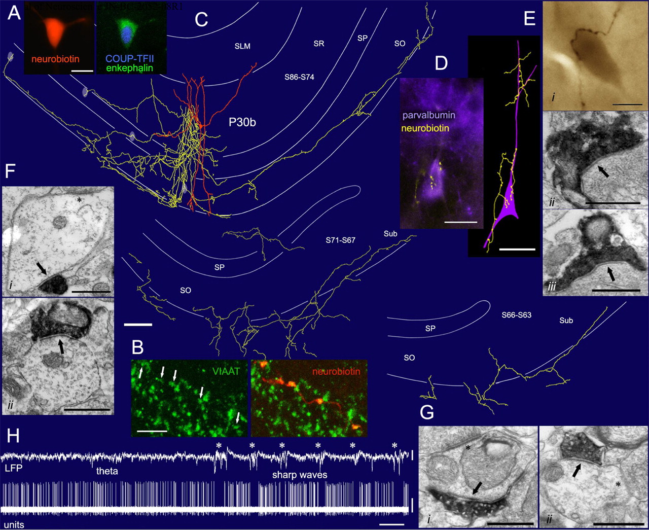
Subicular projection and synaptic output of an identified ENK-expressing interneuron (P30b) recorded in vivo. A, Somatic expression of COUP-TFII (nucleus) and ENK-immunoreactivity. B, VIAAT-immunoreactivity in the axon terminals. C, Reconstruction of the cell. The dendritic arbor (orange) is complete; the axon (yellow) is presented partially in three panels from rostral (section 86) to caudal (section 63) levels. The axon formed a columnar arbor near the soma and an elongated plexus (2.5 mm) in the alveus, continuing into and innervating the subiculum. Four light ellipses depict the location of some synaptically targeted interneurons. D, A parvalbumin-positive interneuron (purple) in stratum pyramidale is selectively innervated on the soma and proximal dendrites by 32 boutons of the interneuron (yellow), as shown in a fluorescent micrograph (left) and a tracing after HRP conversion (right). Ei, A strongly endogenous biotin-containing interneuron in stratum pyramidale is innervated by seven boutons of the identified ENK-expressing cell. Eii, Eiii, Electron micrographs show two boutons making type-II synapses (arrows) on the same soma. Fi, Fii, Electron micrographs show type-II synapses (arrows) onto dendritic shafts of an interneuron (Fi) and a pyramidal cell (Fii) and in stratum radiatum. The asterisk indicates a type-I synapse. Gi, Gii, Electron micrographs show type-II synapses (arrows) onto dendritic shafts of subicular interneurons identified by type-I synapses on the dendritic shafts (asterisks) in these or the following sections. H, Example trace of the LFP (0.3–300 Hz) and spike train (units, 0.8–5 kHz) of the ENK-expressing cell during theta oscillations and sharp waves (asterisks). SO, Stratum oriens; SP, stratum pyramidale; SR, stratum radiatum; SLM, stratum lacunosum moleculare; Sub, subiculum. Scale bars: A, 10 μm; B, 5 μm; C, 100 μm; D–Ei, 20 μm; Eii–G, 0.5 μm. Calibration: H, LFP, 1 mV, 2 s; units, 0.5 mV, 2 s.


