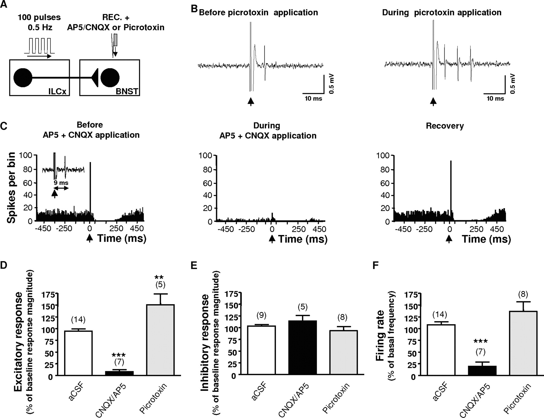
Infusion of glutamatergic receptor antagonists within the BNST block the excitatory drive from the infralimbic cortex on BNST neurons that project to the VTA. A, Diagram of the stimulation protocol: ILCx was stimulated with a 100-pulse train. The CNQX plus AP5 mixture or picrotoxin was microinfused through a pipette adjacent to the recording electrode. B, Effect of local infusion of picrotoxin (1 mm) on a characteristic BNST neuron projecting to the VTA during ILCx electrical stimulation. Representative traces show that picrotoxin enhanced the response probability. C, Effect of the CNQX plus AP5 mixture on a characteristic BNST neuron projecting to the VTA during ILCx electrical stimulation. Typical PSTHs show ILCx-evoked responses before, during, and after (recovery) CNQX plus AP5 injection into the BNST for the same BNST neuron identified as projecting to the VTA. Inset, Orthodromic spikes evoked by stimulation of ILCx. Microinjection of CNQX plus AP5 prevented the short latency activation of BNST neurons evoked by ILCx stimulation, decreased the basal activity, but had no effect on the inhibition. D–F, Graphs illustrating the effects of ionotropic glutamatergic (black bars) and GABA-A (gray bars) antagonists on excitation (D) and inhibition (E) obtained after stimulation of the ILCx and on basal activity (F). Scores are percentages ± SEM of baseline response magnitudes (D, E) or basal activity (F) for BNST neuronal responses evoked by ILCx electrical stimulation during microinjection into the BNST of aCSF (white bars), the mixture of 50 μm CNQX plus 100 μm AP5 (black bars), or 1 mm picrotoxin (gray bars). Numbers of neurons recorded in each group are mentioned in parentheses above each histogram bar. An ANOVA followed by Newman–Keuls test for pairwise comparisons was performed for excitation, inhibition, and basal activity. ***p < 0.001; **p < 0.01. REC, Recording electrode.


